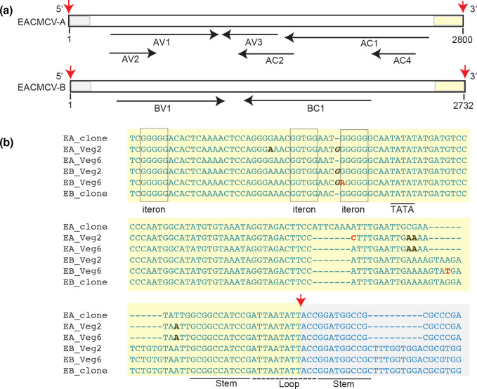
Fig. 6.
EACMCV common regions undergo sequence convergence. (a) Linear maps of EACMCV DNA-A and EACMCV DNA-B. The maps were linearized in the common region at the cleavage site in the top strand of the viral origin of replication. Red arrows mark the 3′-OH and the 5′-P of the nick site [101]. The common region upstream (yellow) and downstream (grey) of the nick site are marked. The open reading frames and directions of transcription are shown by the black arrows below. (b) EACMCV DNA-A and EACMCV DNA-B sequences showing their common regions in the circularized genomic form. The labelling is the same as in (a), with the nick site indicated by a red arrow and the upstream and downstream sequences marked by yellow and grey shading, respectively. The iterons (boxed) and the hairpin motif (stem: underlined; loop: dotted line) involved for the initiation of viral replication are marked. The TATA box (underlined) for complementary sense transcription is also labelled. SNPs showing convergence of the common region sequences of DNA-A and DNA-B are in black typeface, and other SNPs are in red typeface. Insertion is indicated by italics.

