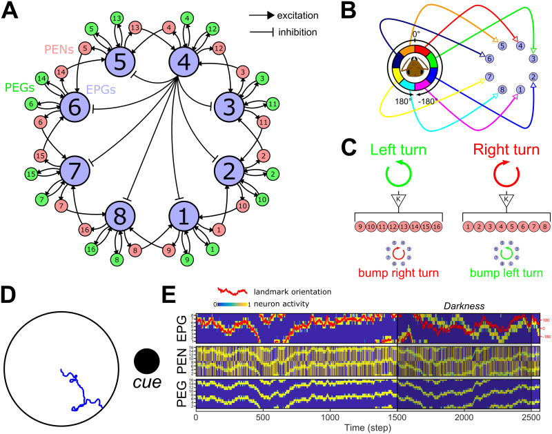
Fig 3. Ellipsoid body compass model.
A. Connectivity diagram between the main neurons of the EB-PB model, EPGs, PEGs and PENs. EPGs have inhibitory connections with each other as indicated, via Delta7 neurons (black lines; one example is shown from the EPG4 in the central part of the diagram). EPGs (light blue) form a recurrent circuit with PEGs (green) while forming connection with neighbouring EPGs through PENs (pink) on each side. B. Connection diagram from the visual circuit to the EPGs. Each angular segment connects in a retinotopic manner to the EPGs. Intrinsically this results in an activity bump forming in the circuit that corresponds to the direction of highest visual contrast. C. Proprioceptive inputs to the PENs. When the agent is engaged in a left turn, PEN1−8 are stimulated, while during a right turn PEN9−16 are stimulated. This will trigger a counter-motion of the bump (posterior view) so that it still indicates the relative direction of the external cue(s). D. Example of the function of the EB model in an arena surrounded by a single cue (here a cylinder represented by the black circle). The blue line shows the path of the agent without any influence of the CX model, i.e. only produced by the steering noise. E. Activity rate of the three neuron types constituting the EB model. On the first line, the activity of EPGs (blue for no activity to yellow for active) shows a perfect following of the cue orientation in the agent’s visual field (red line, scale on the right). Even during a darkness episode (from 1500th to 2500th steps), the model keeps track of the cue position with a relatively low error thanks to the PENs (second line) and PEGs (third line) combined actions.

