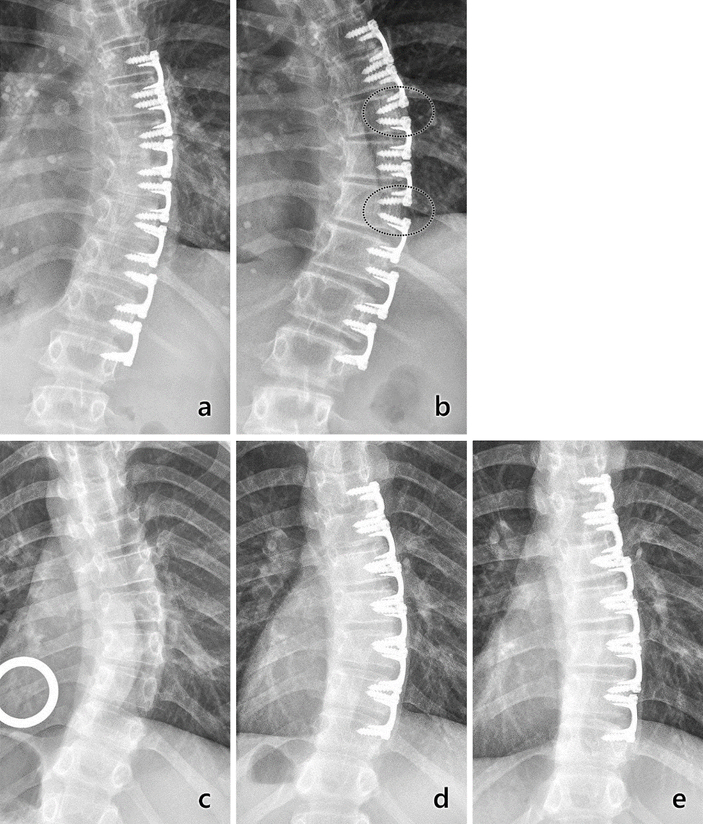
Fig. 8.

Radiographs (PA) of major thoracic curvatures from two subjects, one whose curvature progressed (top) and one whose curvature corrected (bottom). Top: Progression. a) Three-month postoperatively radiograph showing implant placement with 29° curve. b) At 18 months postoperatively, two pairs of adjacent implants clearly moved apart from each other, with apparent bone growth between the implants (circled areas) and increase in curvature. Bottom: Correction. a) Radiograph showing immediately preoperative curvature of 36°. b) Six-month postoperative radiograph showing implant placement and curvature reduction to 25°. c) At 2 years postoperatively, curvature was corrected to 12°. In this case, implants remained tightly grouped.
