. 2021 Sep 17;10:e67670. doi: 10.7554/eLife.67670

Figure 6. Position and presence of glia are not necessary for extracellular vesicle (EV) production and export to occur.

(A) In ttx-1(p767) mutants, the microvilli (Mv) disappeared but TSP-7-wrmScarlet remained enriched in the remaining AFD distal region. Scale bar: 5 μm. (B) Export of TSP-7-wrmScarlet from AFD to AMsh was quantified by counting the number of EVs exported to AMsh. TSP-7-wrmScarlet export is decreased in ttx-1(p767) mutants. Unpaired t-test. (C) In dyf-7(m537) mutants, TSP-7-wrmScarlet remained enriched in AFD receptive endings although the receptive ending was displaced posteriorly in the animal’s head but still embedded within AMsh. TSP-7-wrmScarlet was still exported to AMsh cell body in a similar manner to wild-type controls. Scale bar: 20 μm. (D) Representative images displaying differential tissue capture of EVs when glia is ablated genetically post-embryogenesis. Animals expressed AFD::mEGFP and AFD::TSP-7-wrmScarlet. TSP-7-wrmScarlet is enriched in AFD receptive end in both experimental conditions. EVs are exported to AMsh in control conditions. In the absence of glia, EVs containingTSP-7-wrmScarlet were still produced but were exported to large cells at the surface of the nose, likely the hypodermal cells. TSP-7-wrmScarlet was also exported to amphid sensory neurons. Scale bar: 20 μm for top head images, 5 μm for insets.

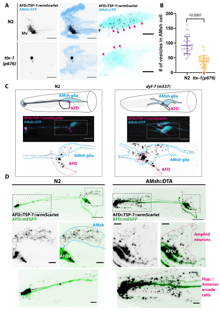
Figure 6.

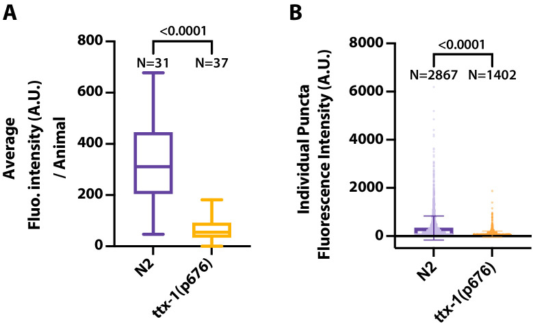
Figure 6—figure supplement 1. In addition to counting extracellular vesicles (EVs) in AMsh, we quantified export of TSP-7-wrmScarlet from AFD to AMsh by extracting the fluorescence of all EVs in AMsh.

Figure 6—figure supplement 1.

(A) The averaged EV fluorescence per animals shows that TSP-7-wrmScarlet export is decreased in ttx-1(p767) mutants. Unpaired t-test with Welch’s correction. (B) The absolute TSP-7-wrmScarlet EV fluorescence per genotype shows an reduced fluorescence intensity of the EV population in ttx-1 mutants. Unpaired t-test with Welch’s correction.
Figure 6—figure supplement 2. Amphid sheath ablation reroutes intake of extracellular vesicles (EVs) to nearby amphid neurons.

Figure 6—figure supplement 2.

(A–D) Representative images of N2 animals expressing AFD::TSP-7-wrmScarlet and pOsm-3::mEGFP, driving expression in a subset of eight amphid neurons (ADF, ADL, ASE, ASG, ASH, ASI, ASJ, ASK). In wild-type animals, all the EVs exported from AFD (magenta arrowheads) end up in AMsh glia (blue dashed outline). AFD was outlined based on expression intensity of TSP-7-wrmScarlet. (E–H) Representative images of AMsh::DTA animals expressing AFD::TSP-7-wrmScarlet and pOsm-3::mEGFP. When glia is ablated, the EVs released from AFD (magenta arrowheads) are rerouted to nearby cells like hypoderm (magenta outline in E) and several amphid neurons. Overlap of TSP-7-wrmScarlet with GFP in the somata of the osm-3 neuronal subset allowed to confirm the presence of TSP-7-wrmScarlet EVs within ADF, ADL, ASE, ASH, ASI, and ASJ (magenta arrowheads in F–H), although we cannot discard the presence of TSP-7-wrmScarlet in non-mEGFP labeled amphid neurons. Scale bar: 20 μm for top head images, 5 μm for insets.