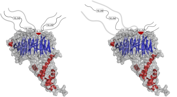
Figure 3.
Allovalency model. An X-ray structure of the SCF E3 ubiquitin ligase components Cdc4 (blue) and Skp1 (red) with the disordered N-terminal region of the yeast cyclin-dependent kinase (CDK) inhibitor Sic1 (cartoon). Multiple phosphorylation sites are positioned in tandem on Sic1 and labelled as SLiMs. Each SLiM is complementary to a binding site on Cdc4 [52]. The dissociation of interaction at one site enables a new interaction at another site. The disordered regions remain flanking as indicated by the blurred lines. PDB ID: 3V7D [52].

