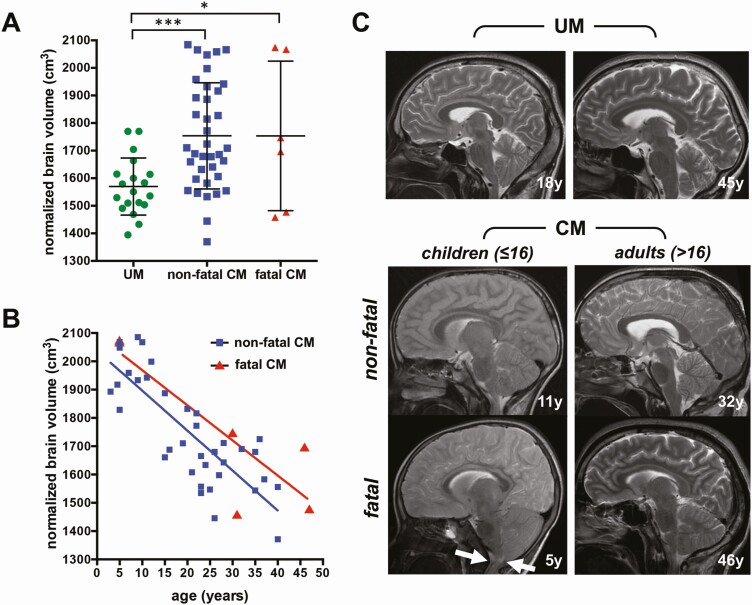
Figure 1.
Comparison of brain volumes on admission between age and disease groups. A, Normalized brain volumes in uncomplicated malaria (UM) and nonfatal and fatal cerebral malaria (CM). B, Correlation between age and normalized brain volume in nonfatal and fatal CM. C, Representative sagittal T2-weighted magnetic resonance imaging of patients with UM (first row) and CM (second and third rows). In pediatric CM patients, the outer cerebrospinal fluid spaces are more reduced due to brain swelling compared with adults. One fatal pediatric CM case showed brain stem herniation with no remaining cerebrospinal fluid space at the craniocervical junction (arrows). *P < .05; ***P < .0005..

