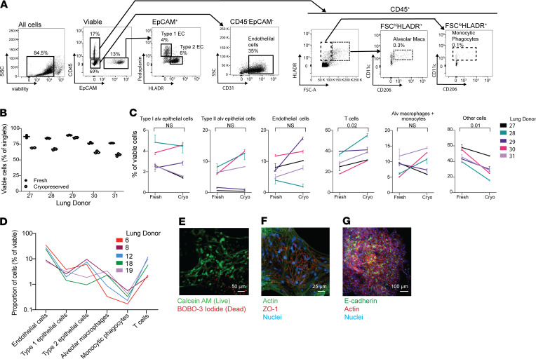
Figure 1. Lung microtissues are viable after cryopreservation.
(A) The composition of cryopreserved lung was assessed by flow cytometry. Measured populations include type I and type II alveolar epithelial cells, endothelial cells, monocytes/macrophages, and T cell populations. (B) Proportion of viable cells in fresh and cryopreserved lung tissue from 5 donors. Samples were run in duplicate before and after cryopreservation. (C) The cellular composition of lung tissue, with a focus on the cell types depicted in A before and after cryopreservation for each of the 5 donors in B. Each line represents the average population present in 2–3 samples, consisting of 20–40 microtissues, from each donor. Error bars indicate variation in the technical replicates for each donor. (D) The cellular composition of cryopreserved samples from an additional 5 donors (separate from those in A and B) was assessed using the gating strategy depicted in C. (E) Viability of cryopreserved tissues was assessed by microscopy using Calcein AM and BOBO-3 iodide in microtissues cultured for 48 hours. Scale bar: 50 μm. (F) Tight junctions in cultured microtissues were also assessed using ZO-1 with costaining for actin and DAPI. Scale bar: 25 μm. (G) Microtissues stained with E-cadherin demonstrate the presence of epithelial cell populations within the cryopreserved samples. Scale bar: 100 μm.

