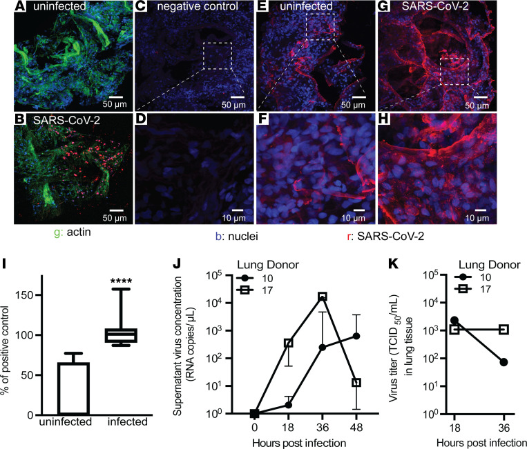
Figure 2. Lung microtissues can be infected with SARS-CoV-2.
(A and B) Micrographs of lung tissue stained with a 1:1 mix of antibodies for spike and nucleocapsid protein to detect SARS-CoV-2 and actin. (C and D) RNA in situ hybridization of SARS-CoV-2–infected lung tissue with a negative control probe specific for the DapB gene of Bacillus subtilis. (E–H) RNA in situ hybridization of lung tissue using a probe specific for the negative strand of the subgenomic E gene of SARS-CoV-2 at 24 hours after infection. Images are representative of microscopy performed on tissue from 2 donors. (I) Quantitative PCR for the subgenomic E gene of SARS-CoV-2 in 16 donors at 24 hours after infection. Range of uninfected samples = 0–77.2, mean = 25.8, standard deviation = 34.6. Range of infected samples = 87.4–157.4, mean = 103.1, standard deviation = 16.7. Significance was determined by the nonparametric Mann-Whitney U test. (J) Copies of SARS-CoV-2 detected in tissue culture supernatant at the indicated time after the start of infection. Samples were infected for 12 hours with 104 PFU and then washed and transferred to fresh media in a new plate. At each time point40 μL aliquots were collected. Error bars are from triplicate wells collected from each donor at each time point. (K) TCID50 assay of lung homogenate using tissue from the same donors as in J at 18 and 36 hours after infection. TCID50 was determined by counting 6 wells per donor at each dilution.

