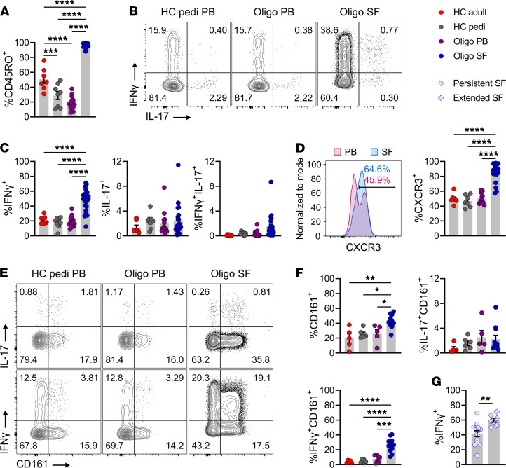
Figure 1. CD4+ memory T cells adopt a Th1 phenotype in the SF of oligo JIA.
(A) Percentage of CD45RO+ cells among CD3+CD4+ lymphocytes. (B) Representative flow staining of cytokine production in stimulated CD3+CD4+CD45RO+ cells (CD4+ Tmem). (C) Percentage of CD4+ Tmem cells expressing IFN-γ, IL-17, or both after stimulation. The PB of adult (n = 7) and pediatric (n = 8) controls and PB (n = 14) and SF (n = 23) of oligo JIA patients were evaluated in A and C. (D) Representative histogram of CXCR3 MFI in paired PB and SF samples from an oligo JIA patient and quantification of CXCR3+ cells among unstimulated CD4+ Tmem cells from HC adult PB (n = 7), HC pediatric PB (n = 7), oligo JIA PB (n = 10), and SF (n = 17). (E) Representative flow staining of CD161 and cytokine production in stimulated cells gated on CD4+ Tmem cells. (F) Percentage of CD161+ cells (unstimulated) and of CD161+ and cytokine dual-expressing cells (stimulated) among CD4+ Tmem cells from HC adult PB (n = 5), HC pediatric PB (n = 6), oligo JIA PB (n = 5), and SF (n = 11). (G) Percentage of CD4+ Tmem cells expressing IFN-γ in SF samples from persistent (n = 14) and extended (n = 7) oligo JIA patients. Summary data on bar graphs are mean ± SEM. *P < 0.05, **P < 0.01, ***P < 0.001, ****P < 0.0001. Statistical testing: (A–F) 1-way ANOVA followed by multiple 2-tailed t tests with Tukey’s correction; (G) 2-tailed t test. HC, healthy control; oligo, oligoarticular juvenile idiopathic arthritis; pedi, pediatric; PB, peripheral blood; SF, synovial fluid.

