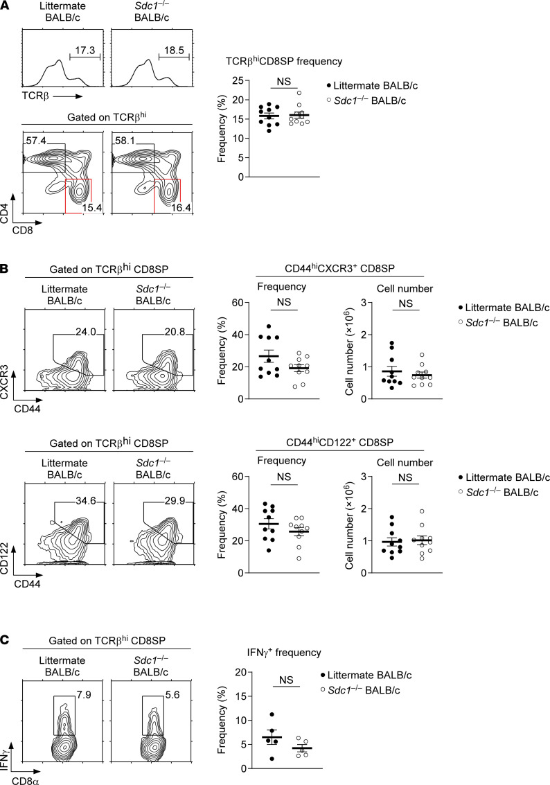
Figure 4. Thymocyte development in CD138-deficient mice.
(A) T cell development in the thymus of Sdc1–/– BALB/c mice. Mature thymocytes were identified by high levels of TCRβ expression and then further assessed for CD4 and CD8 coreceptor expression. Histograms and contour plots (left) are representative, and the graph showing the frequency of CD8 T cells (right) is a summary of 6 independent experiments with a total of 10 Sdc1–/– and 10 WT littermate BALB/c mice. (B) Innate-type marker expression and cell numbers of CD8SP thymocytes of Sdc1–/– BALB/c mice. CD44 versus CXCR3 (top) and CD44 versus CD122 (bottom) expression profiles, and the frequencies and numbers of innate-type cells were assessed in TCRβhi CD8SP thymocytes of Sdc1–/– and WT littermate BALB/c mice. The contour plots represent and the graphs summarize 6 independent experiments with 10 Sdc1–/– and 10 WT littermate BALB/c mice. (C) IFN-γ production by CD8SP cells of Sdc1–/– BALB/c thymocytes. IFN-γ was assessed among TCRβhiCD8SP freshly isolated Sdc1–/– BALB/c thymocytes upon PMA and ionomycin stimulation for 5 hours. Data are representative of 3 independent experiments with a total of 5 Sdc1–/– and 5 WT littermate BALB/c mice. All data are presented as mean ± SEM. P values were determined by unpaired 2-tailed Student’s t test. NS, not significant.

