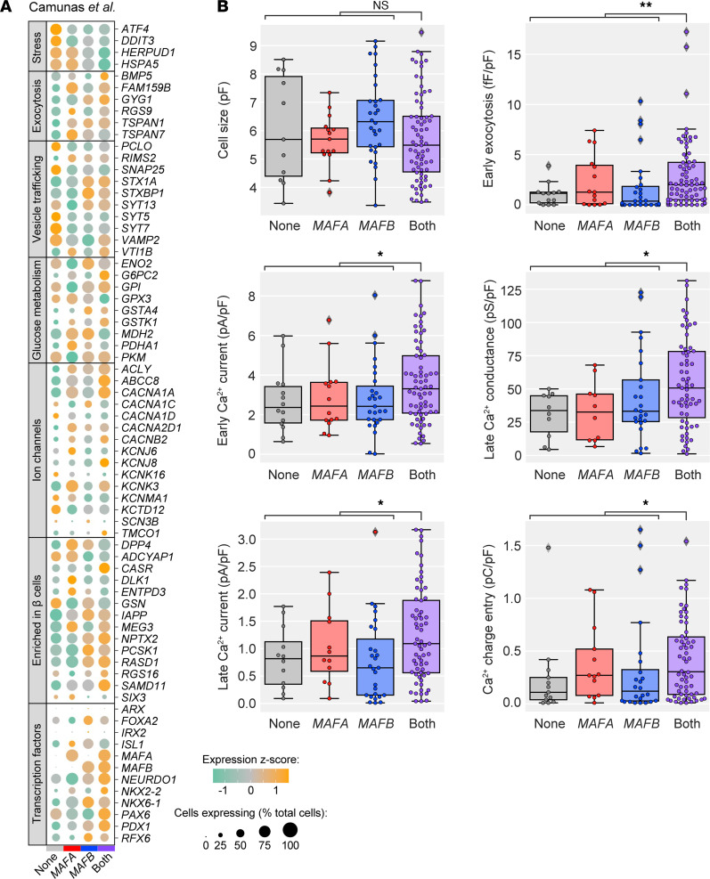
Figure 6. β Cells coexpressing MAFA and MAFB have enhanced electrophysiological activity compared with β cells expressing one or neither factor.
(A) Dot plot showing the relative expression of selected genes in β cells expressing neither MAFA nor MAFB, those expressing only MAFA or only MAFB, and those coexpressing MAFA and MAFB, based on data from Camunas et al. (18). Dot size indicates the percentage of cells with detectable transcripts; color indicates gene’s mean expression z score. (B) Electrophysiological function in MAFA- and MAFB-expressing β cell subpopulations. Significantly higher Ca2+ currents and exocytosis were observed for β cells expressing both MAFA and MAFB with similar cell size across all subpopulations. Mann-Whitney test adjusted for multiple hypothesis testing with Benjamini-Hochberg (BH) procedure; *P < 0.05; **P < 0.01.

