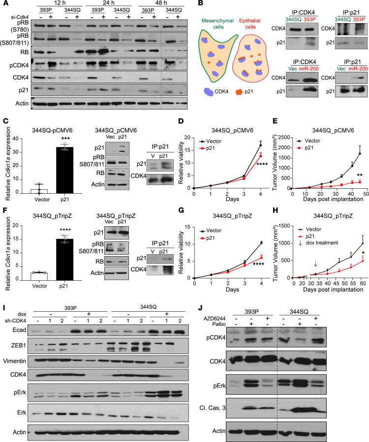
Figure 4. Suppression of p21 in mesenchymal cells regulates CDK4 pathway.
(A) Transient knockdown of CDK4 using 20 nM siRNAs for indicated times followed by Western blot analysis. (B) Graphical representation of the differences in CDK4-p21 complex formation in epithelial and mesenchymal cancer cells. Coimmunoprecipitation (co-IP) of endogenous CDK4 and p21 in epithelial (393P and 344SQ_miR-200) and mesenchymal (344SQ and 344SQ_vec) cell lines. (C) Constitutive overexpression of Cdkn1a in 344SQ cell lines. Relative Cdkn1a mRNA expression, Western blot analysis of CDK4 pathway, and co-IP of CDK4 and p21 in 344SQ cells. (D) Growth rates of 344SQ cells ± p21 constitutive overexpression over 4 days measured by water-soluble tetrazolium salt assay. (E) Tumor volume measurements at indicated time points of 344SQ tumors ± p21 constitutive expression (n = 5 per group). Data are presented as mean ± SEM. (F) Doxycycline-induced overexpression of Cdkn1a in 344SQ cell lines for 48 hours. Relative Cdkn1a mRNA expression, Western blot analysis of CDK4 pathway, and co-IP of CDK4 and p21 in 344SQ cells. (G) Growth rates of 344SQ cells ± p21 overexpression (doxycycline induced) over 4 days measured by WST-1 assay. (H) Tumor volume measurements at indicated time points of 344SQ tumors ± p21 expression with doxycycline feed (n = 9–10 per group). Doxycycline feed was started after tumors reached a size of 100–150 mm3 (indicated by arrow). Data are presented as mean ± SEM. (I) Western blot analysis of 393P and 344SQ cells with CDK4 knockdown for 7 days. (J) Western blot analysis on 393P and 344SQ cells treated with AZD6244 (5 μM) and palbociclib (5 μM) for 48 hours. Data are presented as mean ± SD unless otherwise indicated. Statistical analysis (C, E, F, and H): unpaired 2-tailed Student’s t test and (D and G): 2-way ANOVA test. ****P < 0.0001; ***P < 0.005; **P < 0.001; *P < 0.05.

