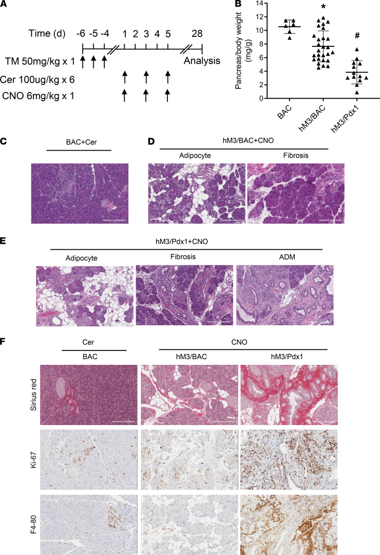
Figure 6. CP develops in hM3R mice.
(A) Three episodes of acute pancreatitis were induced in BAC, hM3/BAC, hM3/Pdx1 mice. The pancreas tissues were harvested at 4 weeks following the initial pancreatitis induction. (B) Pancreas size was measured by pancreas/body weight ratio (mg/g). Mean ± SEM (n ≥ 6). (C) Representative HE staining in cerulein-induced mice. (D) Histology of CNO-induced CP in hM3/BAC mice. (E) In hM3/Pdx1 mice, more severe CP developed. (F) Fibrosis (Sirius red), cell proliferation (Ki-67), and chronic inflammation (F4/80) in BAC, hM3/BAC, and hM3/Pdx1 mice (scale bar: 200 μm). *P 0.05, hM3/BAC+CNO group vs. BAC+Cer group. #P 0.05, hM3/Pdx1+CNO group vs. BAC+Cer group. Two-way ANOVA with Tukey’s test.

