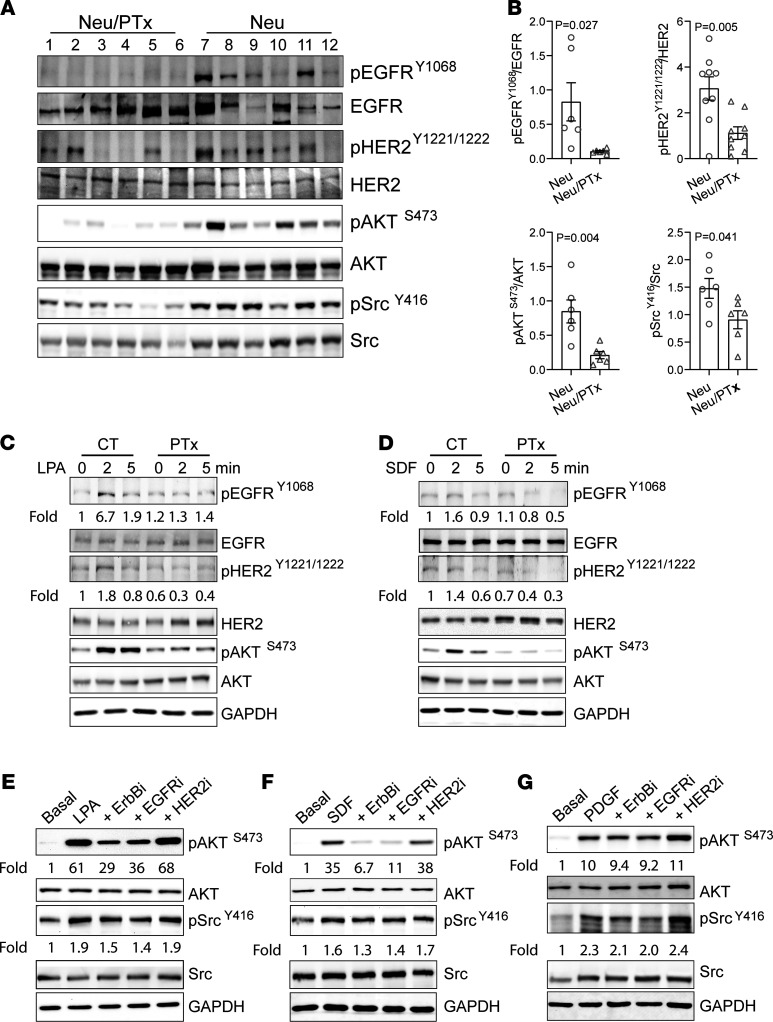
Figure 4. Gi/o-GPCRs induce transactivation of EGFR and HER2 in mammary tumor cells.
(A and B) Western blotting (A) showing decreased phosphorylation of EGFRY1806, HER2Y1221/1222, AKTs473, and SrcY416 in Neu/PTx tumors as compared with Neu tumors. Each lane represents one sample from individual tumors. (B) The Western blot data from A were quantified and expressed as the ratio of the phosphorylated to total proteins. Two-tailed unpaired Student’s t test was used for statistical analysis of the data in B, and P values are shown. (C and D) Western blotting showing phosphorylation of EGFR, HER2, and AKT in Neu cells treated with vehicle control or PTx and stimulated with LPA (C) or SDF1α (D). (E–G) The effect of 1 μM of a pan-ErbB– (sapitinib), EGFR- (erlotinib), or HER2- (cp-724714)specific inhibitor on LPA- (E), SDF1α- (F) or PDGF-stimulated (G) AKT and Src phosphorylation in Neu cells. The phosphorylation of EGFRY1068, HER2Y1221/1222, AKTS473, and SrcY416 was quantified as the ratio of the phosphorylated to total proteins and expressed as the fold increase over basal, which is indicated underneath the images. The images are representatives of at least 3 independent experiments and were assembled from multiple blots run with the samples from the same experiments.

