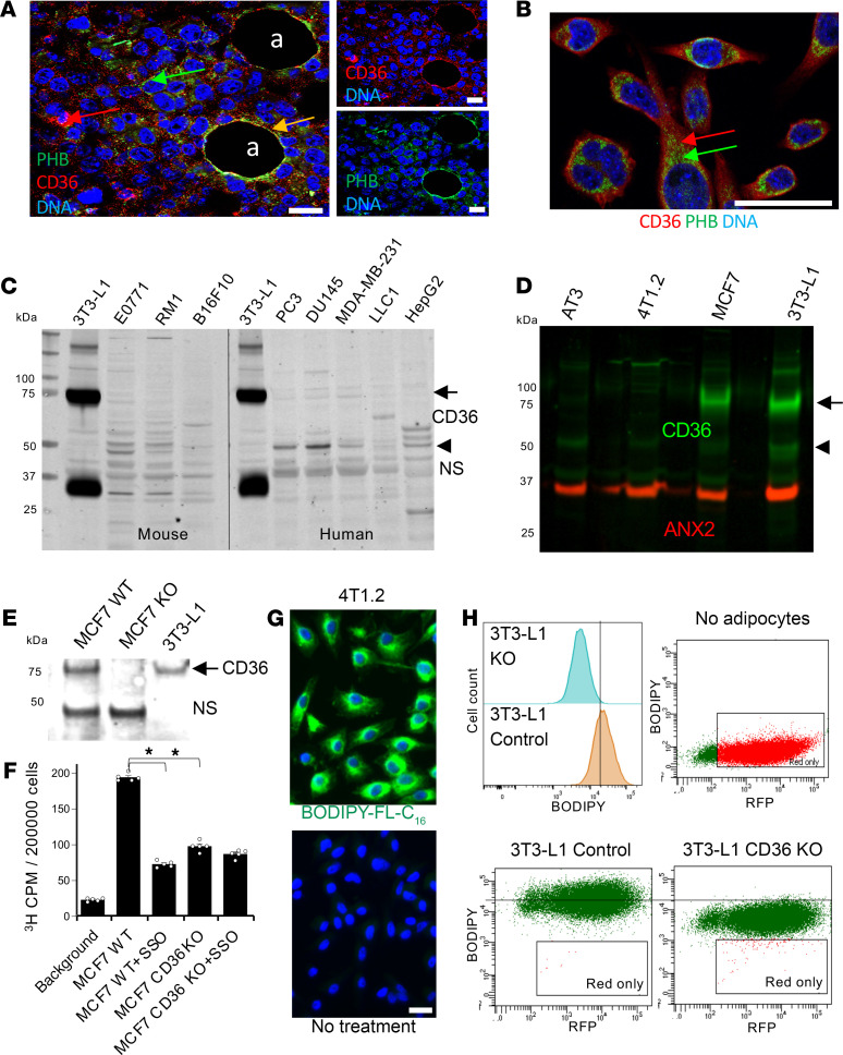
Figure 2. CD36 in cancer cells is not required but can promote LCFA transport.
(A) Immunofluorescence (IF) on paraffin sections of E0771 tumor grafts showing mainly intracellular CD36 and PHB expression in cancer cells; in intratumoral adipocytes (a) colocalization is at the surface (yellow arrow). Blue: nuclei. (B) IF showing that CD36 and PHB (arrows) are intracellular in cultured E0771 cells. Blue: nuclei. (C) Western blotting on extracts from murine and human cell lines demonstrated low expression of CD36 in cancer cells. Arrow: glycosylated CD36. Arrowhead: nonglycosylated CD36. NS, nonspecific band. (D) Western blotting demonstrated expression of CD36 in MCF7 cancer cells comparable to that in 3T3-L1 adipocytes. Arrow: glycosylated CD36. Arrowhead: nonglycosylated CD36. ANX2 immunoblotting: loading control. (E) Western blotting confirming CD36 KO by CRISPR/Cas9 in MCF7 cells transduced with sgCD36. NS, nonspecific band. (F) 3H CPM in indicated cell cultures after 30 minutes exposure to 75 μM 3H-palmitate demonstrated that 3H-palmitate uptake was inhibited by SSO and CD36 KO. n = 5 independent wells. Data are shown as mean ± SEM; *P < 0.01, (1-way ANOVA). (G) 4T1.2 cells preinduced to undergo lipogenesis were untreated or treated with BODIPY-FL-C16 for 10 minutes and imaged for LCFA uptake (arrow). (H) Intercellular fatty acid transfer from 3T3-L1 adipocytes (not plotted) preloaded with BODIPY-FL-C16 (green) to cocultured RFP+ 4T1 cells detected by flow cytometry with 530 nm (BODIPY) and 610 nm (RFP) lasers. The histogram shows the difference in BODIPY-FL-C16 uptake for double-positive (BODIPY-FL-C16+/RFP+) 4T1 cells cocultured with WT versus CD36-KO adipocytes. Scale bar: 50 μm.

