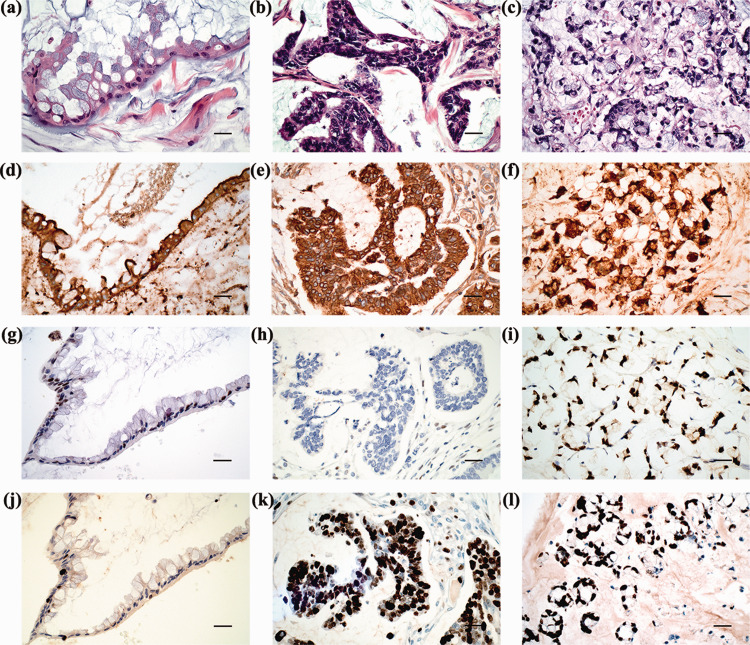
Figure 2.
Representative histopathological features of pseudomyxoma peritonei: histological morphology of low-grade mucinous carcinoma (LGMC) (a), high-grade mucinous carcinoma (HGMC) (b) and high-grade with signet ring cells (HGMC-S) (c) (all haematoxylin and eosin; scale bar 50 µm); strong positive staining for carcinoembryonic antigen in LGMC (d), HGMC (e) and HGMC-S (f); weak focal nuclear staining of p53 in LGMC (g); complete lack of stained tumour nuclei for p53 in HGMC (h); strong positive staining for p53 in HGMC-S (i); low Ki67 LI in LGMC (j); high Ki67 LI in HGMC (k) and HGMC-S (l) (d–l, EnVision staining; scale bar 50 µm). The colour version of this figure is available at: http://imr.sagepub.com.

