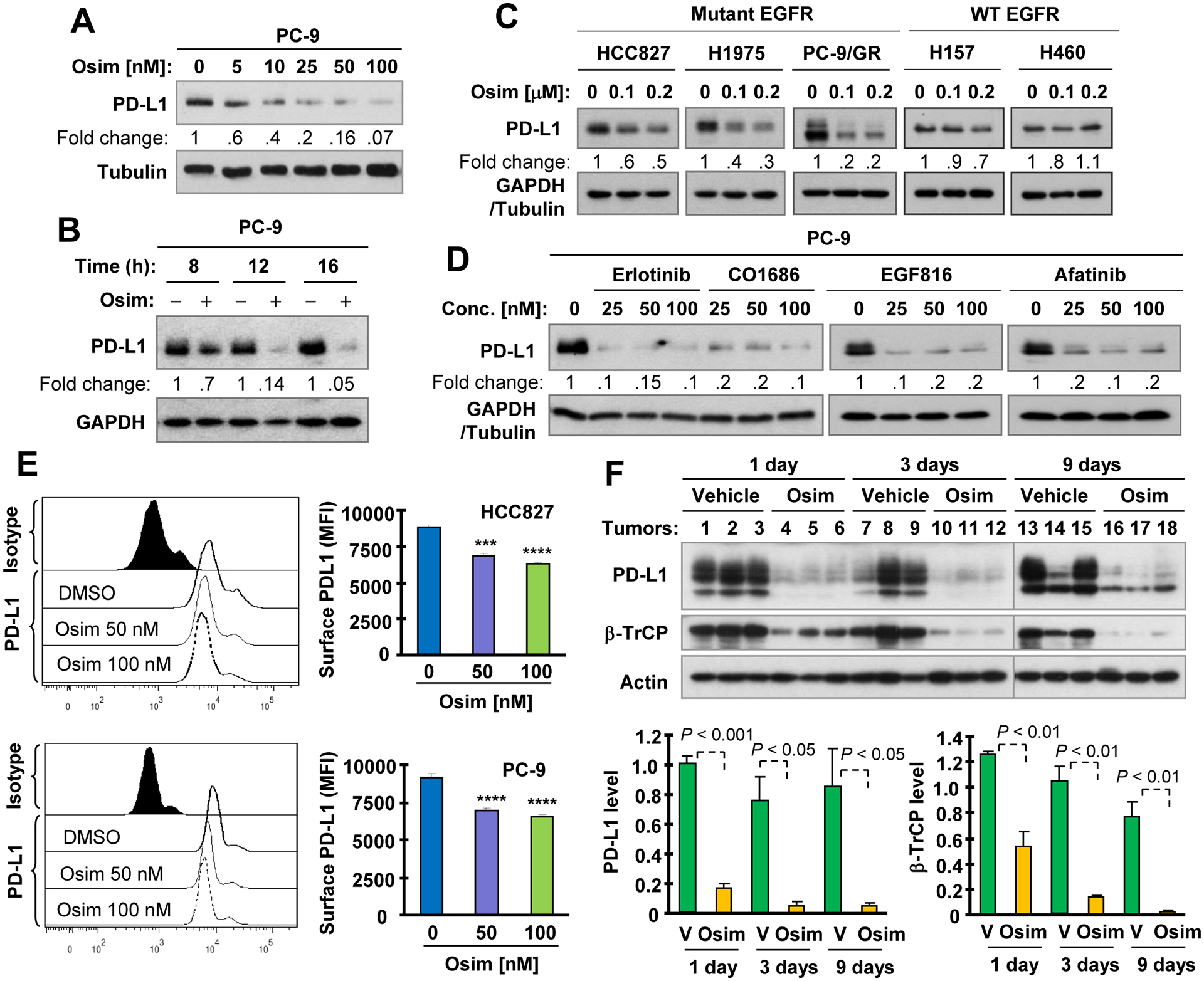
Fig. 1. Osimertinib and other EGFR-TKIs decrease PD-L1 levels in EGFR mutant NSCLC cell lines (A-E) and in xenografts (F).

A-D, The indicated cell lines were exposed to varied concentrations of osimertinib (Osim) for 16 h (A and C), to 100 nM osimertinib for different time as indicated (B) or to different concentrations of EGFR-TKIs as indicated for 16 h (D). Whole cell lysates were then made from these cells and used for detection of PD-L1 and other proteins with Western blotting. E, Both PC-9 and HCC827 cell lines were treated with different concentrations of osimertinib for 36 h. Cell surface PD-L1 was then detected with flow cytometry. ***, P < 0.001 and ****, P < 0.0001 compared with DMSO. F, Whole cell lysates were prepared from PC-9 xenografts treated with osimertinib at 10 mg/kg body weight (og, once/daily) for the indicated times and then subject to Western blotting. Each column is the mean ± SD of triplicate determinations. V, vehicle.
