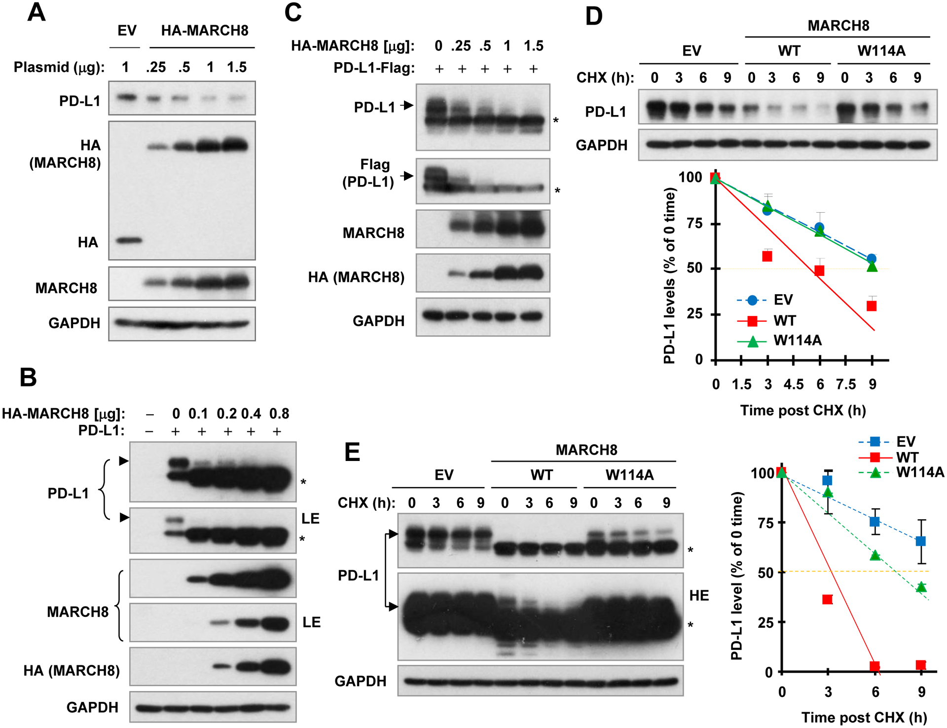
Fig. 3. Enforced expression of ectopic MARCH8 decreases PD-L1 levels (A-C) and enhances PD-L1 degradation (D and E).

A-C, HEK293T cells were transfected with vector control (V) or wild-type (WT) MARCH8 expression plasmid at different amounts as indicated (A), or co-transfected with MARCH8 plus a plasmid expressing non-tagged PD-L1 (B) or flag-tagged PD-L1 (C) for 48 h. D and E, HEK293T cells were transfected with vector, WT or mutated MARCH8 (W114A) for 40 h or co-transfected with MARCH8 and PD-L1 (non-tagged) expression plasmids for 24 h, followed by addition of 10 μg/ml CHX for varied time as indicated. Whole cell lysates were then prepared from the above treatments for Western blot analysis to detect the indicated proteins. PD-L1 levels were plotted relative to those at time 0 of CHX treatment after being quantified by NIH Image J software and normalized to GAPDH (D and E). LE, low exposure; HE, high exposure; *, uncharacterized band generated by ectopic expression of PD-L1.
