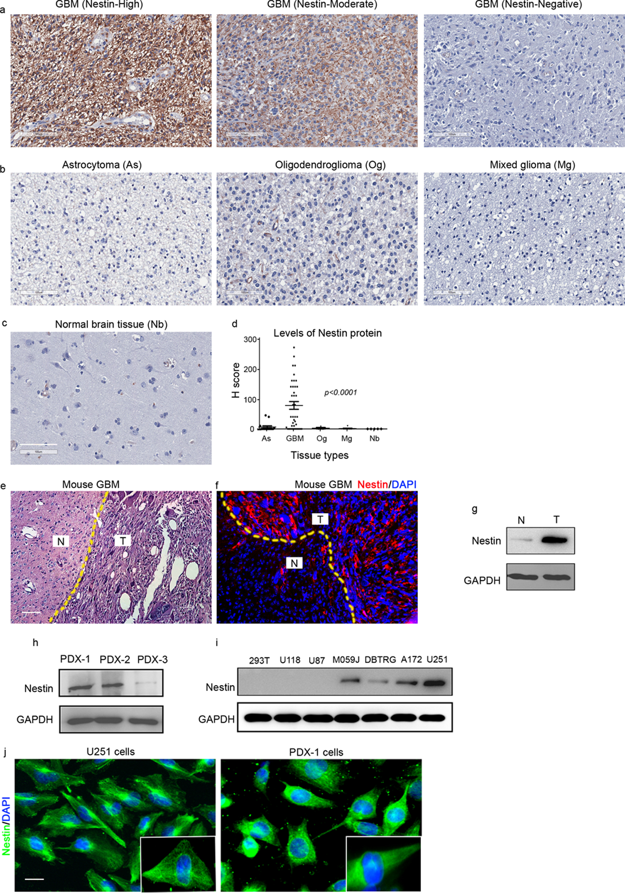
Figure 1. Nestin is highly expressed in GBM cells.

a-d, Representative images of immunohistochemical staining of Nestin from TMA cores of GBM specimens with high, moderate or negative expression of Nestin (a); from astrocytoma (As), oligodendroglioma (Og) or mixed glioma (Mg) (b), as well as from adjacent normal brain tissue (Nb) (c). The levels of Nestin proteins in TMA cores were compared by H score (d), p<0.0001, one way ANOVA. scale bar: 100 μm
e-g, HE staining (e) and immunostaining of Nestin (f) from a brain from a GBM mouse model. DAPI was used to counterstain cell nuclei. The abundance of Nestin protein in tumor tissue and normal brain tissue was examined by western blotting. N, adjacent normal brain tissue; T, GBM tumor tissue. scale bar: 50 μm
h-i, Nestin protein in tumor cells from 3 GBM PDX lines (h) and available GBM cell lines (i) was examined by western blotting. GAPDH was used as a loading control.
j, Nestin protein in U251 cells and PDX-1 cells was examined by immunocytochemistry. Insets show the magnified images of intracellular distribution of Nestin. scale bar: 20 μm.
