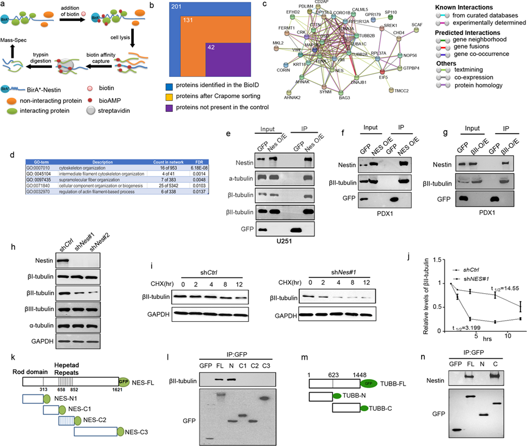
Figure 4. Nestin stabilizes βII-tubulin in GBM cells by physical interaction.

a, A schematic representation of the BioID approach to identify Nestin-interacting proteins.
b, Sorting process of the proteins identified by the Nestin BioID assay. 201 proteins were identified by MS, 70 proteins were excluded based on CRAPome analysis and 89 proteins were present in the control U251 cells, resulting in 42 proteins identified by the BioID assay as Nestin-interacting proteins in U251 cells.
c, PPI network of Nestin interacting proteins (revealed by the BioID approach) based on STRING-based protein interacting networks.
d, Top 5 terms based on GO enrichment analyses of Nestin interacting proteins.
e, Whole cell extracts from U251 cells virally infected with a Nestin-GFP vector or an empty GFP vector, were immunoprecipitated using an antibody against GFP. GFP, α-tubulin, βI and βII-tubulins, Nestin proteins were detected in the input (whole cell extracts) and immunoprecipitates by western blotting.
f–g, PDX-1 cells were infected with a lentivirus carrying a GFP-tagged vector encoding Nestin (f) or βII-tubulin (g), or an empty vector as a control. Whole cell lysates from PDX-1 cells were immunoprecipitated using an antibody against GFP (f and g). Nestin, βII-tubulin and GFP were detected in the input and immunoprecipitates by western blotting.
h, U251 cells were virally infected with Nestin shRNA (shNes#1, shNes#2) or scrambled shRNA (shCtrl). 96hrs following the infection, U251 cells were harvested for examination of Nestin, α-, βI-, βII- and βIII-tubulin proteins by western blotting. GAPDH was used as a loading control.
i–j, Nestin-deficient U251 cells and control U251 cells were treated with CHX for 12hrs and collected at designated timepoints to examine the levels of βII-tubulin by western blotting (i). The relative levels of βII-tubulin in U251 cells (normalized to the level at 0hr) were quantified based on the densitometry of the western blot band. The half-life of βII-tubulin in U251 cells was calculated by Prism 7.
k–l, Schematic representation of the GFP-tagged Nestin fragments (k). U251 cells were trans fected with full-length or fragments of Nestin. The cell lysate was immunoprecipitated with an antibody against GFP, and examined for βII-tubulin and GFP by western blotting (l).
m–n, Schematic representation of the GFP-tagged βII-tubulin fragments (m). The cell lysate of U251 cells transfected with the full-length or fragments of βII-tubulin, was immunoprecipitated with an antibody against GFP, and examined for Nestin and GFP by western blotting (n).
