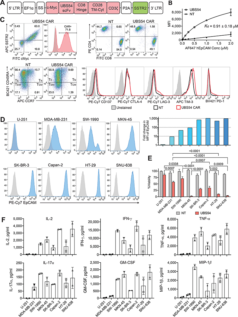
Figure 1. The in vitro activity of EpCAM CAR T cells against tumor cell lines.
A, Schematic representation of the lentiviral vector encoding EpCAM-specific UBS54 CAR. The expression of the CAR construct is driven by the EF1α promoter, and SSTR2 is placed following a ribosome-skipping sequence P2A. A c-Myc tag was introduced at the N-terminus for CAR detection. SS, signal sequence; scFv, scFv derived from UBS54 monoclonal antibody; CD28 TM-Cyt, CD28 transmembrane and cytosolic domains. B, The affinity of the UBS54 CAR was determined by staining CAR-expressing Jurkat T cells with serially diluted EpCAM. Kd values were calculated using the one-site nonlinear regression model. Data represent mean±s.d. (n=3). NT, non-transduced T cells. C, Representative flow plots showing CAR expression, T-cell phenotypes (CD4, CD8, TN CCR7+CD45RA+, TCM CCR7+CD45RA−, TEM CCR7−CD45RA−, TE CCR7−CD45RA+), and activation and exhaustion markers (CD137, CTLA-4, LAG-3, TIM-3, and PD-1) in CAR T cells and NT. D, Surface expression of EpCAM in tumor cell lines was determined by flow cytometry. EpCAM densities in tumor cell lines were presented as fold-increases in EpCAM MFI normalized to unstained cells. E, Cytolytic activity of CAR T cells against EpCAM-expressing target cells and EpCAM-negative control U-251 cells. CAR T cells were co-incubated with target cells at an E:T ratio of 1:1, and 24 hours later, the cell viability of target cells after CAR T-cell or NT treatment was normalized to that of the no T cell control. Data represent mean±s.d. (n=3). Statistical significance was determined by unpaired, two-tailed t-test. F, Cytokines in culture supernatants were measured after co-incubation of CAR T cells and target cells for 24 hours (n=2).

