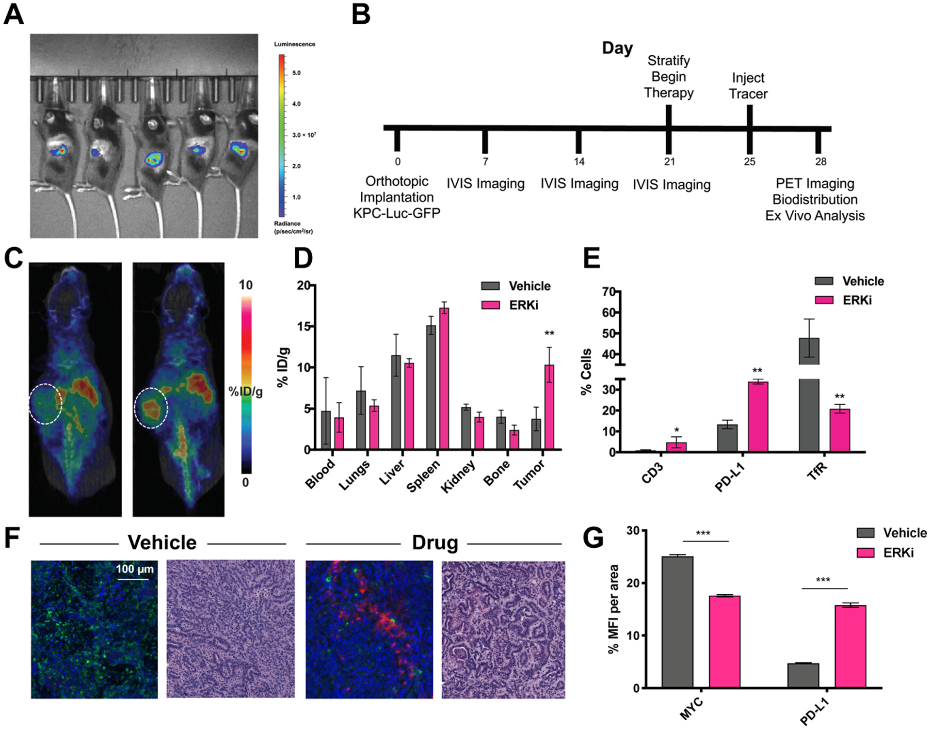
Figure 4. ERK inhibition increased uptake of [89Zr]Zr-DFO-anti-PD-L1 in orthotopic KPC-Luc-GFP mice.

A. Bioluminescent imaging of KPC-Luc-GFP tumors 2 weeks post-orthotopic injection. B. Timeline of implantation and drugging. Animals received ERK inhibitor SCH772984 once daily at 90 mg/kg for 7 days. C. Uptake of [89Zr]Zr-DFO-anti-PD-L1 increase upon ERK inhibition in vivo and is quantified via ex vivo biodistribution (D). E. Ex vivo flow cytometry of KPC-Luc-GFO tumors shows an increase in PD-L1 expression an CD3+ T cells correlated to a decrease in TfR expression. **P < 0.01. F. Immunofluorescent analysis of MYC (green) and PD-L1 (pink) expression ex vivo in KPC tumors and H&E stains of serial sections. G. Quantification of immunofluorescent analysis of MYC+ and PD-L1+ cells in KPC PDAC tumors. Abbreviation: MFI: mean fluorescent intensity.
