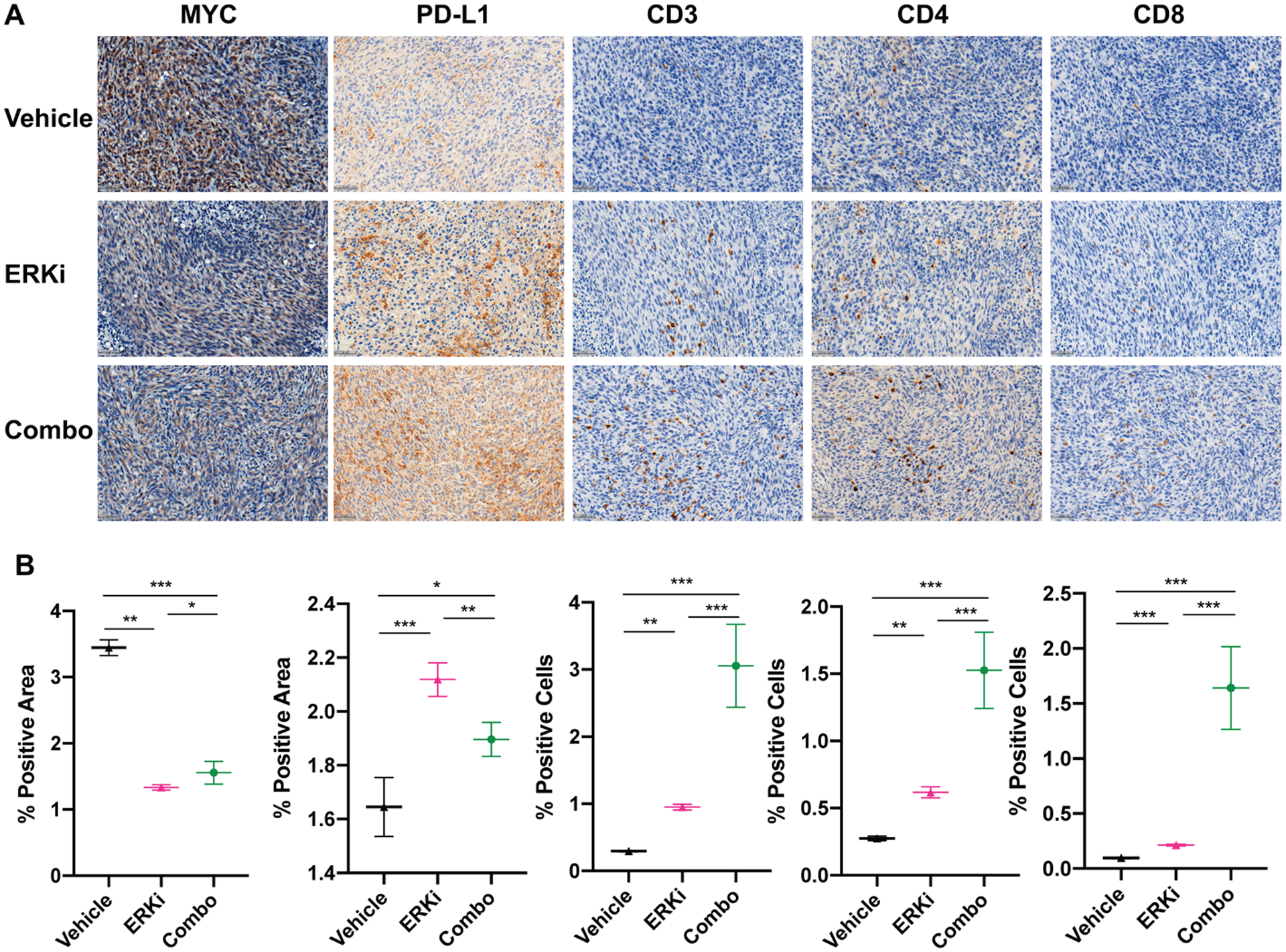
Figure 6. Immunohistochemical analysis of ex vivo tissues collected at single endpoint indicate a potential T-cell mediated mechanism.

A. Representative images of serial sections from the same tumors stained for MYC, PD-L1, CD3, CD4, and CD8 with trends quantified in (B). Significant trends are observed along with decreases in MYC staining and increase in PD-L1 staining (along with CD3, CD4 and CD8) in each treatment arm compared to respective controls. *P < 0.05, **P < 0.01, ***P < 0.001.
