. 2021 Aug 10;20(10):1904–1915. doi: 10.1158/1535-7163.MCT-20-0638

Figure 3.

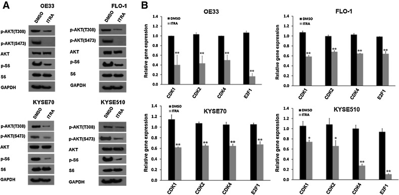
Figure 3. Itraconazole inhibits AKT activity in both EAC and ESCC cell lines. A, Western blot analysis of p-AKT (Thr308 and Ser473), AKT, p-S6, and S6 expression in OE33, FLO-1, KYSE70, and KYSE510 cells treated with 2.5 μmol/L itraconazole or DMSO for 48 hours. GAPDH is used as a loading control. B, RT–qPCR analysis of CDK1, CDK2, CDK4, and E2F1 genes in OE33, FLO-1, KYSE70, and KYSE510 cells treated with 2.5 μmol/L itraconazole or DMSO for 48 hours. Values represent the mean fold change ± SEM for three experiments relative to GAPDH. Black bars, DMSO; gray bars, itraconazole. *, P < 0.05; **, P < 0.01 vs. DMSO control by Student t test.

Itraconazole inhibits AKT activity in both EAC and ESCC cell lines. A, Western blot analysis of p-AKT (Thr308 and Ser473), AKT, p-S6, and S6 expression in OE33, FLO-1, KYSE70, and KYSE510 cells treated with 2.5 μmol/L itraconazole or DMSO for 48 hours. GAPDH is used as a loading control. B, RT–qPCR analysis of CDK1, CDK2, CDK4, and E2F1 genes in OE33, FLO-1, KYSE70, and KYSE510 cells treated with 2.5 μmol/L itraconazole or DMSO for 48 hours. Values represent the mean fold change ± SEM for three experiments relative to GAPDH. Black bars, DMSO; gray bars, itraconazole. *, P < 0.05; **, P < 0.01 vs. DMSO control by Student t test.