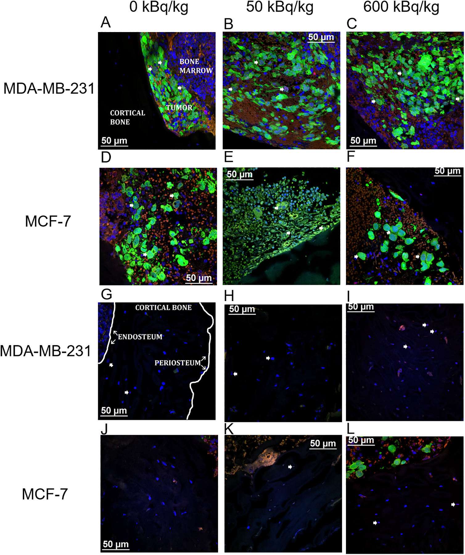
Figure 1. Representative confocal microscopic images of transverse tibial bone marrow sections containing human breast cancer cells that were inoculated into animals pre-treated with 223Ra dichloride and assayed for γ-H2AX formation.

MDA-MB-231 (A–C) and MCF-7 (D–F) labeled with CellTracker™ Green CMFDA and stained with anti-γ-H2AX antibody (AlexaFluor™ 568 red) to visualize DNA damage. The tumor, bone marrow, and cortical bone have all been demarcated (A). Nuclear counter-staining with DAPI (blue) visualizes DNA damage in mouse osteocytes in the tibiae of animals inoculated with MDA-MB-231 cells (G–I) and MCF-7 cells (J–L). The inner (endosteum) and outer (periosteum) cell layers surrounding the cortical bone have been noted (G). Arrowheads delineate γ-H2AX positive cells. Images acquired with a Nikon A1R microscope with CFI Apochromat TIRF 60XC oil (NA 1.40), DS-Fi3 camera and NIS-Elements C software.
