
Figure 5. Quantification of apoptosis in transverse tibial bone marrow sections stained with TUNEL from mice inoculated with human breast cancer cells.
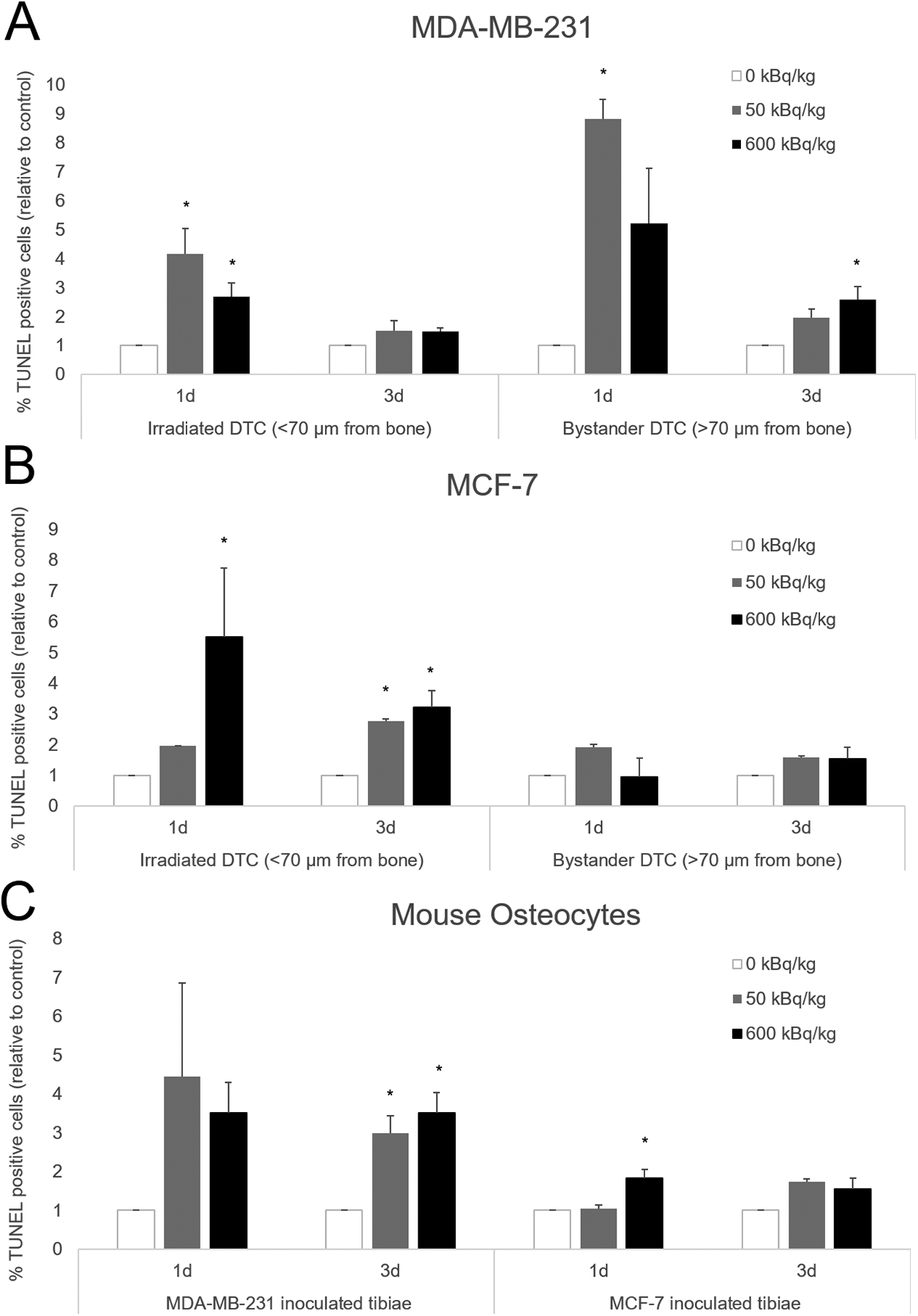
MDA-MB-231 (A), MCF-7 human breast cancer cells (B), as well as mouse osteocytes (C) from both groups. Mice were euthanized 1- and 3-days following cell inoculation after previously being treated with 50 or 600 kBq/kg of 223RaCl2. Control mice were given saline. Irradiated disseminated tumor cells were demarcated from bystander disseminated tumor cells by being less than or greater than 70 μm from the inner surface of the cortical bone. The percentage of TUNEL positive disseminated tumor cells and mouse osteocytes were determined from 1–3 sections for each animal with n=2–7 per group. That percentage of TUNEL positive cells was then normalized to a percentage of TUNEL positive cells from a control animal that had their tissue processed and embedded at the same time. Errors bars represent Standard Error of the Mean (SEM) with asterisks denoting significance (see Supplementary Table S6).