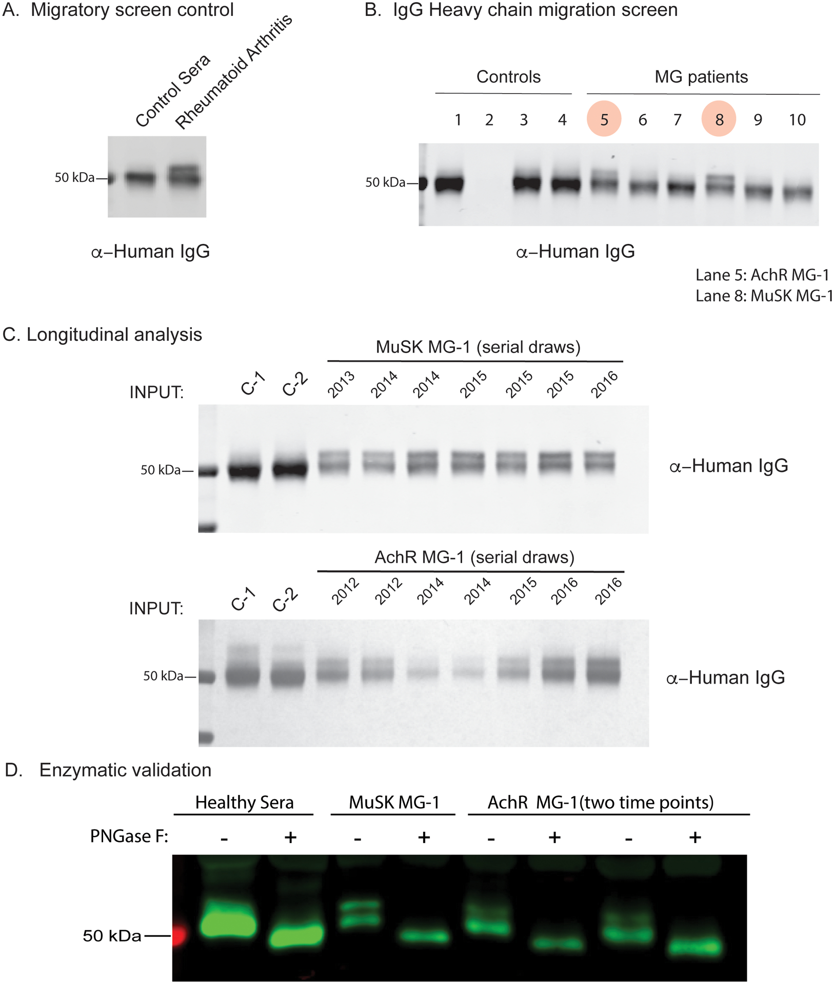
Figure 2. Proteomic analysis of total serum IgG glycosylation suggests elevated Fab N-linked glycosylation in MG.

Total serum IgG heavy chain migratory patterns from SDS-PAGE and immunoblotting with anti-human secondary are shown for sub-panels. (A) Serum IgG heavy chain migration pattern from non-inflammatory control and a patient with RA shown by immunoblot. (B) Total serum IgG heavy chain migration patterns for non-inflammatory controls (Lane 1, 3, 4), A/G beads only (No antibody, Lane 2) and AChR MG (Lanes 5, 6, 7) or MuSK MG (Lanes 8, 9, 10) shown by immunoblot. Lane 5 corresponds to subject AChR MG-1 and lane 8 corresponds to subject MuSK MG-1. (C) Longitudinal serum IgG heavy chain patterns from subjects MuSK MG-1 and AChR MG-1 shown by immunoblotting with an anti-human IgG secondary antibody. (D) Enzymatic validation of N-linked glycosylation in total circulating IgG from a healthy individual, MuSK MG-1 and AChR MG-1 (two distinct serial draws), shown by immunoblot. Human IgG was incubated with or without PNGase F then subject to SDS-PAGE and immunoblotted with an anti-human IgG secondary antibody. Treatment with PNGase F results in loss of migration phenotype. RA = Rheumatoid Arthritis.
