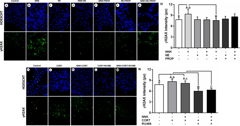
Figure 4.
Exposure to the NNK and norepinephrine and γH2AX nuclear expression in oral keratinocytes. (A) Control. (B) NNK. (C) Norepinephrine. (D) NNK and norepinephrine. (E) NNK and propranolol. (F) Norepinephrine and propranolol. (G) NNK, norepinephrine and propranolol. (H) NNK increased the γH2AX nuclear expression levels. Beta-blocker propranolol inhibited the increased DNA damage caused by the carcinogen. Cortisol increased the γH2AX nuclear expression in oral epithelial cells. (I) Control. (J) Cortisol. (K) NNK and cortisol. (L) Cortisol and RU486. (M) NNK, cortisol and RU486. (N) Cortisol increased the γH2AX nuclear expression levels in the presence or absence of carcinogen NNK. Glucocorticoid receptor blocker RU486 inhibited the increased DNA damage caused by the hormone. The results are expressed as the mean ± standard deviation (SD). Upper- and lower cases equal letters indicate a statistically significant difference (p < 0.0001). NNK = 4 (N-metil-N-nitrosamine)-1-(3-piridil)-butano-1-one). NE = norepinephrine. PROp = propranolol. CORT = cortisol. NNK, NE, PROP and RU486 were used at 10 µM. CORT was used at 100 nM. NOK-SI cells were exposed to NNK and/or stress hormones in the presence or absence of their antagonists for 72 h.

