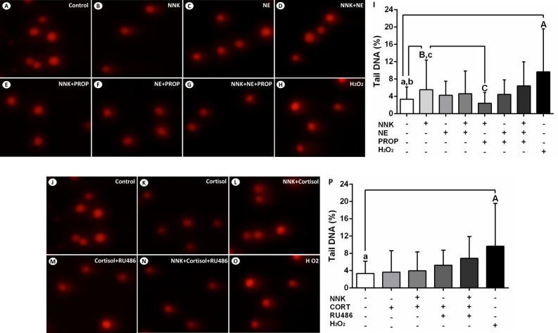
Figure 5.
NNK promotes the oxidation of basis in the DNA of NOK-SI cells. (A) Control. (B) NNK. (C) Norepinephrine. (D) NNK and norepinephrine. (E) NNK and propranolol. (F) Norepinephrine and propranolol. (G) NNK, norepinephrine and propranolol. (H) H2O2 (positive control). (I) Cells treated with H2O2 or NNK exhibited an increase in the DNA damage, when compared to untreated cells after exposure to FPG enzyme. Beta-adrenergic antagonist propranolol blocked this effect. Norepinephrine in the presence or absence of NNK did not induce significant oxidation of basis. Cortisol did not promote significant oxidative DNA damage. (J) Control. (K) Cortisol. (L) NNK and cortisol. (M) Cortisol e RU486. (N) NNK, cortisol and RU486. (O) H2O2 (positive control). (P) H2O2 promoted DNA damage in oral keratinocytes. Cells treated with cortisol alone or combined with NNK did not show significant changes regarding the oxidation of basis. The results are expressed as the mean ± standard deviation (SD). Upper- and lower cases equal letters indicate a statistically significant difference (p < 0.0001). H2O2 = hydrogen peroxide. NNK = 4 (N-metil-N-nitrosamine)-1-(3-piridil)-butano-1-one). NE = norepinephrine. PROp = propranolol. CORT = cortisol. NNK, NE, PROP and RU486 were used at 10 µM. CORT was used at 100 nM. NOK-SI cells were exposed to NNK and/or stress hormones in the presence or absence of their antagonists for 72 h.

