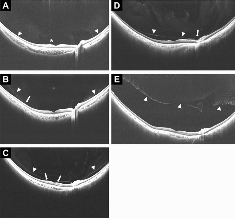
Figure 3.

PVD stages observed on widefield SS-OCT. The right eye of a 5-year-old girl with stage 0 PVD shows the paramacular bursa (asterisk) without separation of the posterior vitreous cortex and the retina (A). The right eye of a 23-year-old man with stage 1 PVD shows the partial separation of the posterior vitreous cortex that did not extend into the fovea (arrow) (PLM-VD of 4876 µm (≥ 750 µm) (B). The right eye of a 47-year-old man with stage 2 PVD shows vitreoretinal separation from a part of the fovea with persistent attachment to the foveola (arrows). This is defined as a shorter (nasal) PLM-VD of 136 µm (< 750 µm) (C). The right eye of a 69-year-old man with stage 3 PVD shows separation from the entire macula with persistent attachment to the optic nerve (arrow) (D). The right eye of an 88-year-old woman with stage 4 shows no adhesion between the retina and the posterior vitreous cortex. Arrowheads in all images indicate the posterior vitreous (E).
