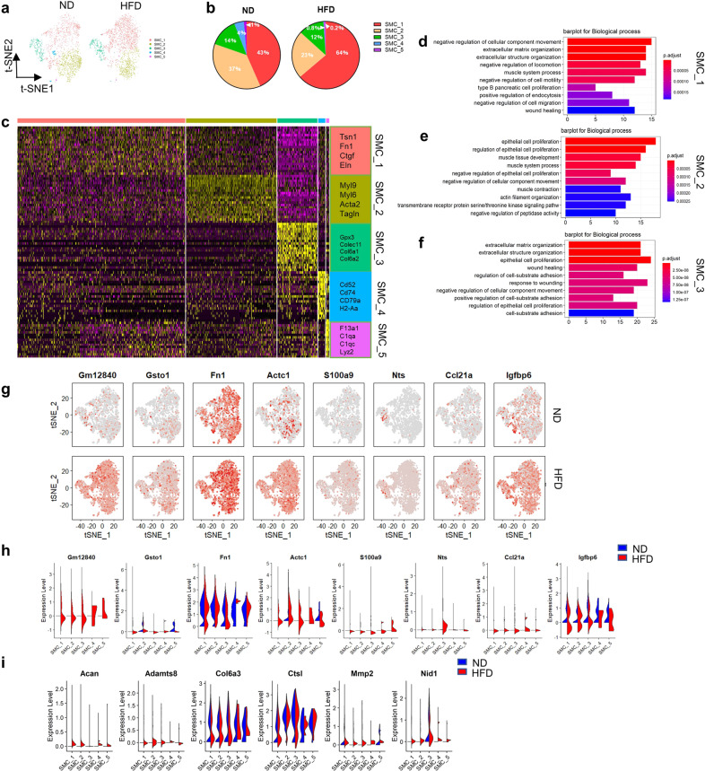
Fig. 3. Comparison of SMC subpopulations in ascending aortas from ND and HFD mice.
a, t-SNE plots of the SMC subpopulations (SMC_1, SMC_2, SMC_3, SMC_4, and SMC_5) from the ND (2,601 cells) and HFD (2,370 cells) groups. b, Percentages of the SMC subpopulations from the ND (SMC_1, 43%; SMC_2, 37%; SMC_3, 14%; SMC_4, 4%; and SMC_5, 1%) and HFD (SMC_1, 64%; SMC_2, 23%; SMC_3, 12%; SMC_4, 0.8%; and SMC_5, 0.2%) groups. c, Heatmap of the top 20 marker genes per subpopulation. d–f, Top 10 pathways associated with the SMC_1 (d), SMC_2 (e), and SMC_3 (f) clusters. g, Feature plots of the expression of selected marker genes for the SMC subpopulations from the ND and HFD groups. h, Violin plots of the expression of selected marker genes for the SMC subpopulations from the ND and HFD groups. i, Expression of extracellular matrix (ECM) degradation-associated genes for SMC subpopulations as shown by violin plots.

