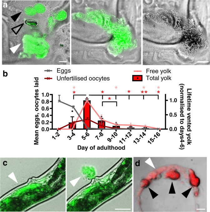
Fig. 1. Post-reproductive C. elegans hermaphrodites vent yolk.
a Vented yolk pools and unfertilised oocytes on culture plates from hermaphrodites on d4 of adulthood, expressing vit-2::GFP. For other examples and separate images for Nomarski and epifluorescence microscopy see Supplementary Fig. 1c. Scale 50 μm. b Lifetime reproductive schedule, oocyte production, and proportion of total vented yolk (from oocytes + free yolk) and free yolk quantitated from VIT-2::GFP on plates and normalised to days 4–6. Trails were started at the L4 stage, and day 1 as specified denotes the first 24 h after the L4 stage, which includes part of day 1 of adulthood. Mean ± S.E.M. of 3 trials displayed (n = 50 worms per trial for venting and 10 per trial for brood sizes). *P < 0.05, **P < 0.01 by one-way ANOVA (Tukey correction; statistical tests performed on raw data), compared to day 4–6; red, total yolk (free + oocytes); pink, free yolk alone. Total yolk left to right compared to d5-6 for stars P = 0.010, 0.027, 0.024, 0.004, 0.011; d7–8 vs 9–10 P = 0.047. Free yolk left to right P = 0.016, 0.019, 0.017, 0.017. c Yolk vented through the vulva of a day 4 adult. Live imaging of VIT-2::GFP day 4 adults performed with yolk initially present in the uterus (left) and then seen vented 7 sec later (right) (white arrowhead). For presence of yolk in the uterus, other examples of yolk venting, time series, and comparison to egg laying on day 2 of adulthood see Supplementary Fig. 1d–f and Supplementary Movie 1. Scale 50 μm. d Lipid in vented yolk and unfertilised oocytes from day 4 adults subjected to vital staining with the lipid dye Bodipy 493/503. Scale 50 μm. White arrowhead: yolk pools, black arrowhead: unfertilised oocytes, and open arrowhead: egg.

