
FIGURE 1: Representative Colonic Polyps from Patient 1 (1A) and Patient 3 (1B).
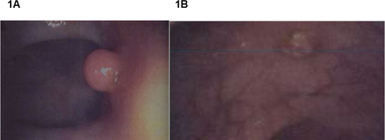
1A) A pedunculated juvenile polyp found in the ascending colon of Patient 1. 1B): A sessile polyp found in the ascending colon of Patient 3. Although these lesions are located in the same portion of the colon, the precancerous potential of the sessile polyp in Patient 3 is more concerning.