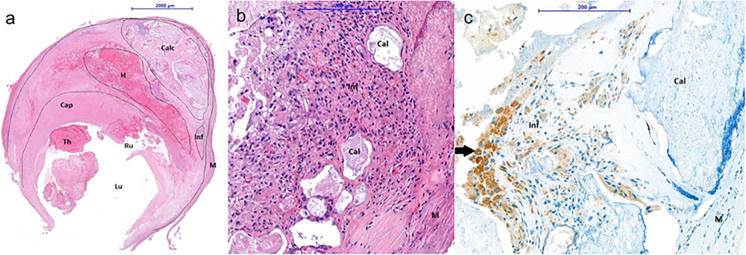
Fig. 1.
Representative cross-section of an unstable carotid plaque removed during carotid endarterectomy stained by HE. The plaque is composed of a large amount of fibrotic tissue in the plaque and the cap of the plaque. Grade 4 plaques are characterized by a rupture (Rup) in the fibrous cap of the plaque (Cap), presence of a thrombus (Thr) and intraplaque hemorrhage (Hem); inflammation (Inf); calcification (Cal); medial smooth muscle cells (M); lumen (Lum) (a). A representative magnified image of inflammatory cells plaque tissue stained by HE (b). An image with CD68 positive macrophage staining. Arrows points to positively stained cells in plaque tissue. The high number of CD68 positive macrophages and foam cells in the plaques are histological features of the unstable plaques (c) [51].

