Figure 1.

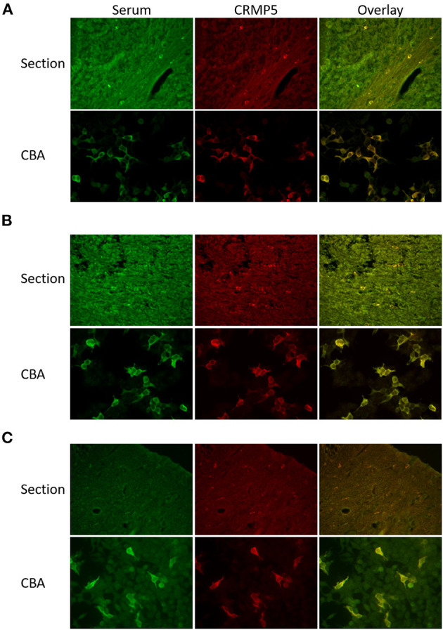
Comparison of CRMP5 staining patterns in cerebellar sections and CRMP5-CBA. (A) Section: Serum from a patient with thymoma and myasthenia gravis stains oligodendrocytes in cerebellar white matter (green). A rabbit anti-CRMP5 antibody (red) stains the same cells. Overlay seen in yellow. CBA: Patient serum (green) specifically detect CRMP5-transfekted HEK293 cells. Anti-CRMP5 antibody (red) is used to detect CRMP5 positive cells. Overlay seen in yellow. (B) Section: Serum from a patient with peripheral neuropathy, lung cancer, Hu, Zic4, and CRMP5 antibodies. The CRMP5 signal is masked in the additional staining of the other antibodies in the patient serum (green). Co-staining with rabbit anti-CRMP5 antibody (red). The overlay shows that the serum also detects CRMP5 positive oligodendrocytes. CBA: The serum (green) stains CRMP5 positive cells in the CRMP5-CBA. Rabbit anti-CRMP5 (red) is used to verify CRMP5 positive cells. Overlay seen in yellow. (C) Serum from a patient with lung cancer and peripheral neuropathy (green) is negative in cerebellar sections.
