FIGURE 5.
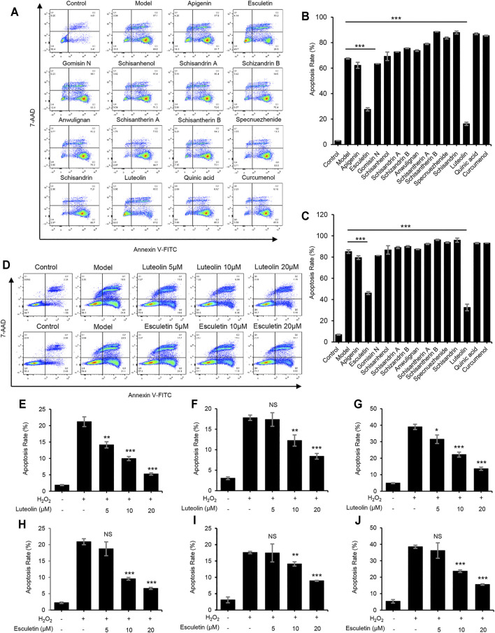
Luteolin and esculetin suppress H2O2-induced apoptosis. (A) Apoptosis of L02 cells treated with apigenin, esculetin, gomisin N, schisanhenol, schisandrin A, schisandrin B, anwulignan, schisantherin A, schisantherin B, specnuezhenide, schisandrin, luteolin, quinic acid, and curcumenol (40 μM) and then exposed to APAP, as detected by flow cytometry. (B) The percentage of early apoptotic cells from samples described in A. (C) The percentage of total apoptotic cells from samples described in A. (D) Apoptosis of L02 cells treated with esculetin or luteolin (5, 10, and 20 μM) and then exposed to APAP, as detected by flow cytometry. (E–G) The percentage of early apoptotic cells (E), late apoptotic cells (F), and total apoptotic cells (G) treated with luteolin (5, 10, and 20 μM). (H–J) The percentage of early apoptotic cells (H), late apoptotic cells (I), and total apoptotic cells (J) treated with esculetin (5, 10, and 20 μM). Data are represented as the mean ± SD using biological samples. The significance of the differences was analyzed using unpaired Student’s t-test: *p < 0.05, **p < 0.01, ***p < 0.001 vs. the control, NS, not significant.

