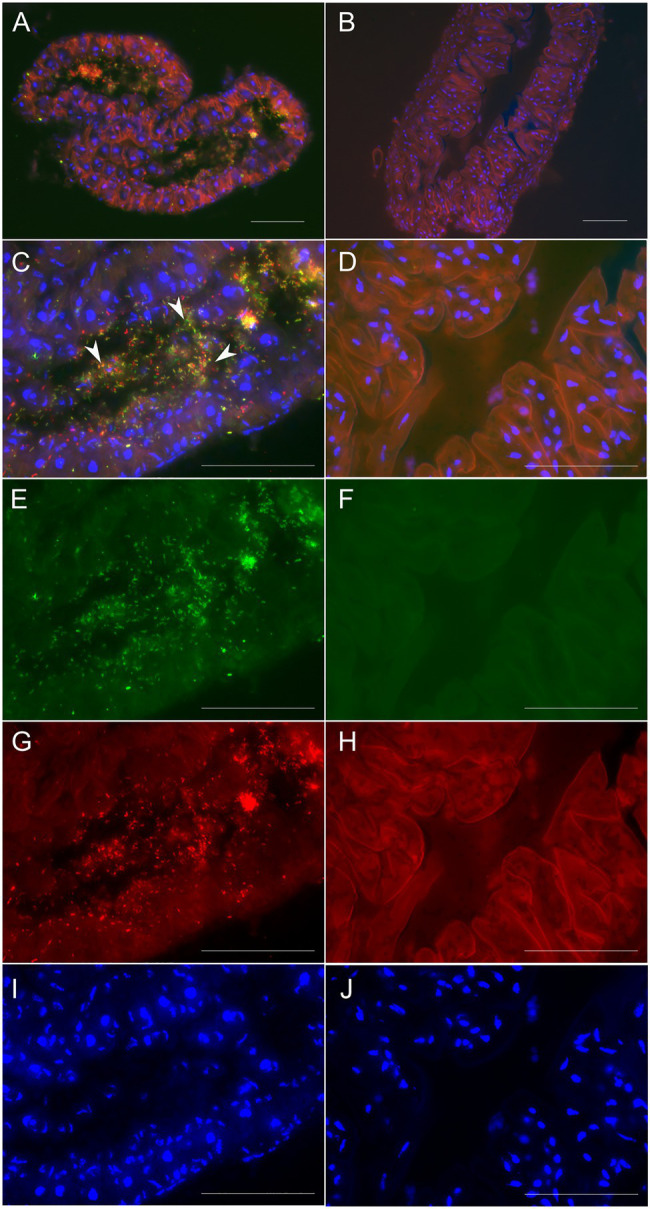
Figure 5.

Micrographs showing environmental acquisition of B. gladioli Lv-StA by L. villosa or absence in an unexposed control. (A,C,E,G,I) Lv-StA presence in cross-sections (8μm) of the accessory glands of an adult L. villosa that had been exposed to Lv-StA during the larval stage. (B,D,F,H,J) show an aposymbiotic control individual. In (A) and (C), symbiont cells are represented in yellow and white arrowheads denote exemplary symbiont cells. Fluorescent signals in individual channels correspond to (E,F) Burkholderia specific labeling in green (Burk_16S-Cy5), (G,H) general eubacteria labelled in red (EUB338-Cy3), and (A–D,I,J) nucleic acids unspecifically labeled in blue (DAPI) showing host cell nuclei. The scale bars represent 50μm. For clarity, H shows autofluorescence from the host tissues in the Cy3 channel.
