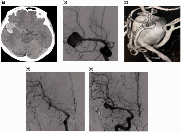
Figure 2.
(a–e) (A) patient with Fisher grade 3/Hunt & Hess grade 2 subarachnoid hemorrhage. Non-Contrast CT image show (a) a large localized clot adjacent to the aneurysm. DSA and 3D image (b and c) show a complex wide-neck aneurysm at the right MCA bifurcation. Immediate angiography (d) shows RR1 total occlusion of the aneurysm after a YSAC (Neuroform Atlas) procedure. 1-year follow-up angiography shows (e) RR3 recanalization of the aneurysm.

