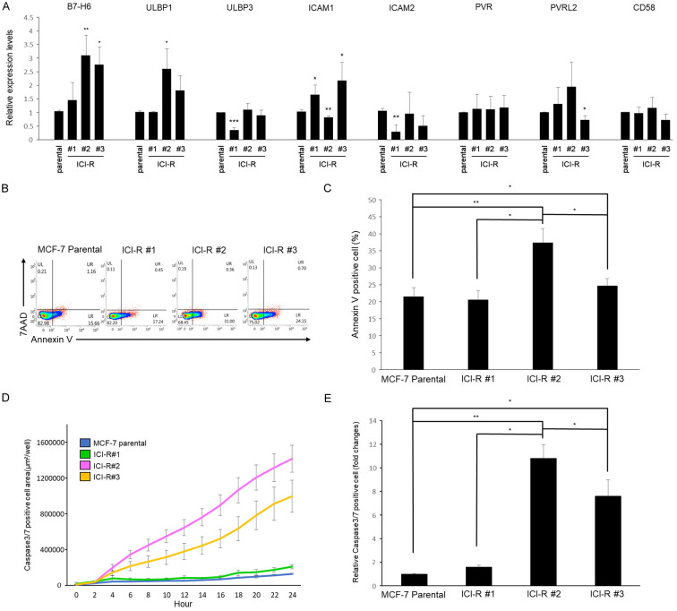
Figure 2.
Gene expression of cell surface molecules involved in innate immunity and cytotoxicity of NK cells. A. mRNA expression of the indicated cell surface genes was analyzed by qRT-PCR normalized to GAPDH. Results are presented as mean ± SD based on three independent experiments. The statistical significance was assessed by t-test. *, P < 0.05, **, P < 0.01. B. MCF7 and the ICI-R cells were co-cultured with NK-92 cells for 24 h. Apoptotic cells were analyzed by gating with annexin V and 7-AAD running through flow cytometry. C. The quantitative results of cytotoxicity assay by flow cytometry. Results are presented as mean ± SD based on three independent experiments. The statistical significance was assessed by t-test. *, P < 0.05, **, P < 0.01. D. Real time detection of cytotoxicity of NK92 cells co-cultured with MCF-7 and ICI-R cells. Results are presented as mean ± SD based on three independent experiments. E. The quantitative results of cytotoxicity assay by IncuCyte. Results are presented as mean ± SD based on three independent experiments. The statistical significance was assessed by t-test. *, P < 0.05, **, P < 0.01.

