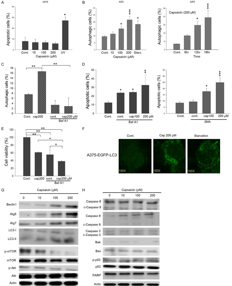
Figure 2.
Capsaicin favorably triggers autophagy but not apoptosis in A375 melanoma cells. A. Cells were treated with capsaicin or ethanol for 24 hours. The percentage of apoptotic cells was evaluated by flow-cytometry, and the results are presented as the percentage of apoptotic cells. Values (means ± SDs) are from at least three independent experiments (*P<0.05). B. Cells were exposed to various concentrations of capsaicin/ethanol for 15 hours or 200 μM of capsaicin for different time length. Autophagy was determined by AO staining using flow cytometry, analysis and the results are expressed as a percentage of autophagic cells. Values (means ± SDs) are from at least three independent experiments (*P<0.05, ***P<0.001). C. Cells were pre-treated with 10 nM Baf A1 for 1 h before exposed to capsaicin for 15 h. Autophagy was determined by AO staining using flow cytometry, analysis and the results are expressed as a percentage of autophagic cells. Values (means ± SDs) are from at least three independent experiments (**P<0.01). D. Cells were pretreated with 10 nM Baf A1 (left panel) or 10 μM 3-MA (right panel) for 1 h before treated with capsaicin or ethanol for 24 hours. The percentage of apoptotic cells was determined by flow cytometry, and the results are expressed as a percentage of apoptotic cells. Values (means ± SDs) are from at least three independent experiments (*P<0.05, **P<0.01, ***P<0.001). E. Cells were pre-treated with 10 nM Baf A1 for 1 h before exposed to various concentrations of capsaicin for 24 h and cell viability was determined by MTS-based assays. Values (means ± SDs) are from no less than three independent experiments. There was a significant difference observed in cell viability in experimental groups as opposed to the controls (*P<0.05, **P<0.01). F. A375-EGFP-LC3 cells were serum starved or treated with capsaicin or ethanol for 15 hours. EGFP fluorescence was observed under a fluorescence microscopy. G, H. A375 cells were exposed to capsaicin or ethanol for various concentrations and aliquots of cell lysates were resolved by SDS-PAGE and analyzed for protein expression by Western blotting. β-actin was used as an internal loading control to monitor for equal loading. Representative images are provided from at least three independent experiments.

