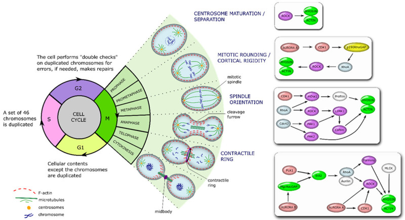
Figure 2.

The regulation of actin dynamics during different phases of mitosis. The scheme shows different phases of the cell cycle, depicting the intracellular processes that take place during mitotic (M) phase, and placing a special emphasis on signalling cascades that regulate actin cytoskeleton during the cell cycle progression. During prophase chromosomes condense, spindle fibers emerge from the centrosomes, nuclear envelope breaks down, and centrosomes move towards the opposite poles of the cell. Then, in prometaphase, kinetochores arise at the centrosomes and mitotic spindle microtubules attach to the kinetochores. Next, during metaphase, chromosomes line up at the metaphase plate, while each sister chromatid gets attached to a spindle fiber, outgrowing from the opposite poles. During anaphase, centromere split in two and sister chromatids are pulled towards the opposite poles, while spindle fibers start to elongate the cell. This is followed by telophase, where chromosomes at the opposite poles begin to decondense, and nuclear envelope, surrounding each set of chromosomes, starts to reform. Then, spindle fibers continue to push poles to the opposite directions. Finally, a cleavage furrow, which separates newly formed daughter cells, is formed. The scheme on a right depicts functional relation of proteins, participating in a control of actin dynamics during the M-phase of the cell cycle.
