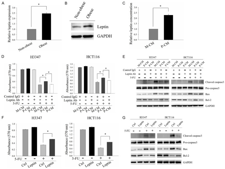
Figure 2.
Leptin was involved in obese adipocyte-induced 5-FU resistance in CRC cells. (A and B) mRNA and protein levels of leptin in non-obese and obese adipocytes were analyzed by quantitative RT-PCR (A) and Western blot analysis (B), respectively. (C) Amounts of leptin in M-CM and P-CM were measured by ELISA. (D and E) H3347 and HCT116 cells were pre-incubated with M-CM or P-CM with/without 2 μg/ml leptin neutralizing antibodies for 48 hours followed by 5-FU treatment for 48 hours. Cell viability was evaluated by MTT analysis (D). The protein expressions of apoptosis-related molecules, cleaved caspase3, Bax and Bcl-2, were analyzed by Western blot analysis (E). (F and G) H3347 and HCT116 cells pre-treated with 100 ng/ml leptin recombinant protein for 48 hours were subjected to 5-FU treatment for another 48 hours. MTT assay and Western blot analysis were used to detect cell viability (F) and the expressions of apoptosis-related molecules (G), respectively. M-CM, non-obese adipocyte-derived conditioned media. P-CM, obese adipocyte-derived conditioned media. GAPDH served as the loading control. Data are expressed as the mean ± SEM. SEM, error bars. *P<0.05 by Student’s t test or one-way ANOVA followed by Bonferroni’s post hoc test.

