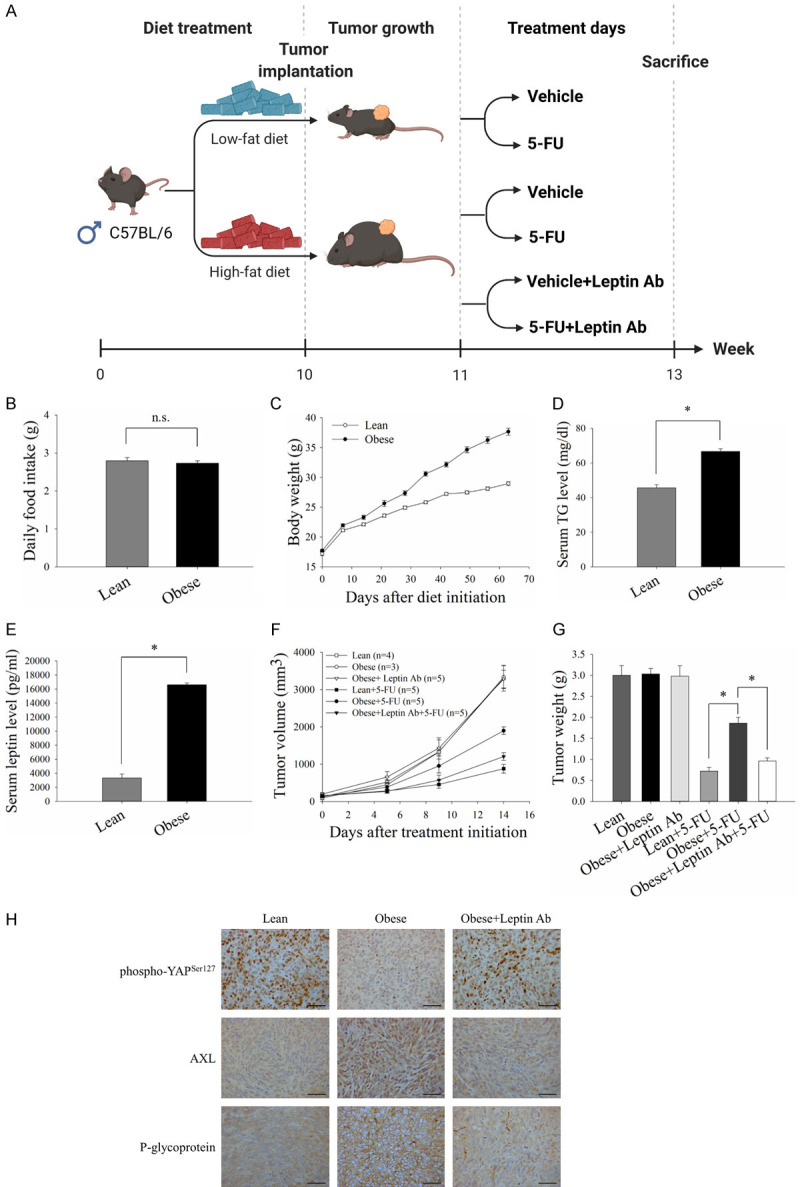
Figure 7.

Leptin inhibition sensitized CRC cells to 5-FU in obese mice. C57BL/6 mice were fed on a HFD or LFD from 5 weeks of age until the end of the experiments. MC-38 cells (1×10⁶) were subcutaneously implanted 10 weeks after initiating the diet, and treatment was started when the tumor burden reach about 100 mm3. 5-FU (25 mg/kg) and neutralizing antibodies against leptin (50 μg per mouse) were injected intraperitoneally into tumor-bearing mice for 2 weeks. Goat IgG antibody was served as the control. The tumor volume was calculated using the formula: V = (width2 × length)/2. A. Timeline of the animal experiments and treatment schedule for the tumor-bearing mice. B and C. Food intake and body weight changes of the mice fed on a HFD or LFD. D. Triglyceride (TG) levels in mice serum were detected using a TG colorimetric assay. E. ELISA was used to measure the serum level of leptin in the mice. F and G. Average tumor growth curves and tumor weights of CRC tumors in the mice with the indicated treatments. Tumor growth was monitored every 3-4 days. H. Representative immunohistochemistry images for phospho-YAP (Ser127), AXL and P-gp staining in CRC tumor sections (Scale bar, 50 μm). Data are expressed as the mean ± SEM of at least three mice in each group. SEM, error bars. n.s., not significant. *P<0.05 by Student’s t test or one-way ANOVA followed by Bonferroni’s post hoc test.
