
Fig. 1. Schematic of radiogenomics platform for DCE MRI-based tissue procurement and genomic analysis.
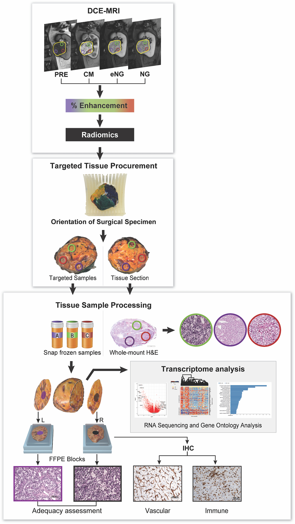
Patients underwent magnetic resonance imaging (MRI), including dynamic contrast-enhanced (DCE) MRI, for vascularity determination. Yellow circle delineates the entire tumor. Other colored circles represent the targeted regions of interest (tROIs) corresponding to areas of high and low enhancement on the DCE MRI. After surgical resection, each tumor was anatomically co-registered and sliced to match the imaging plane. Tissues samples from the same location in the tumor specimen and approximate size as the tROIs were collected as targeted samples. These samples were snap frozen and further processed for RNA extraction, and genomic analysis as indicated. The results of the gene expression analysis were then correlated with the MRI-derived measures. Immunohistochemistry (IHC) slides were generated from flanking sections of each targeted tissue sample for assessment of vascular and immune features. Tumors were also graded based on the International Society of Urological Pathology (ISUP) grading system. Overall, 45 out of 49 primary tumors had more than one tROI: 16 tumors had 2 tROIs, 17 tumors had 3 tROIs, 7 tumors had 4 tROIs, 3 tumors had 5 tROIs, 1 tumor had 6 tROIs and 1 tumor had 8 tROIs.