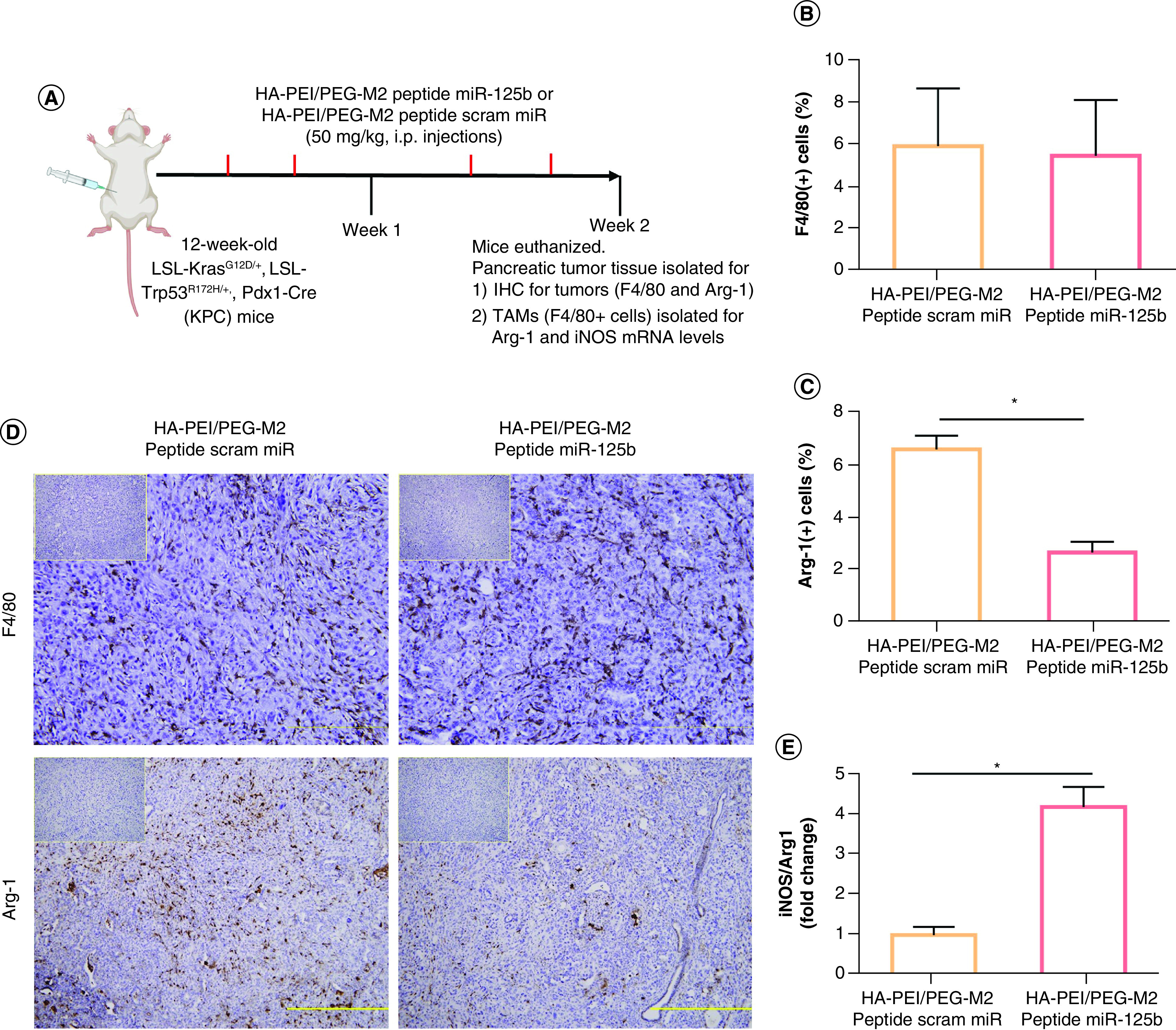
Figure 5. . In vivo polarization of tumor-associated macrophages using miR-125b encapsulated HA-PEI/HA-PEG-peptide in KPC mice.

(A) Schematic method for the in vivo macrophage repolarization study in KPC mice. (B) F4/80 and (C) Arg1 immunostaining were performed on KPC tumor sections and photographs were taken at ×20 (for F4/80) and ×10 (for Arg-1) magnification. (D) The consecutive section was stained with isotype IgG as a negative staining control and is shown in the upper right corner. Results are expressed as a percent of F4/80+ or Arg1+ cells ± SD per ×20 or ×10 field, respectively. (E) The ratio of iNOS/Arg1 in KPC tumor macrophages treated with HA-PEI nanoformulations. qPCR was used for quantification of the gene expression level with β-actin as the internal control.
*Significant compared with the control group; p < 0.01.
HA-PEG: Hyaluronic acid-poly(ethyleneglycol); HA-PEI: Hyaluronic acid-polyethyleneimine; IHC: Immunohistochemistry; KPC: KrasLSL.G12D/+; p53R172H/+; PdxCretg/+; M2: Alternatively activated macrophage; TAM: Tumor associated macrophage.
