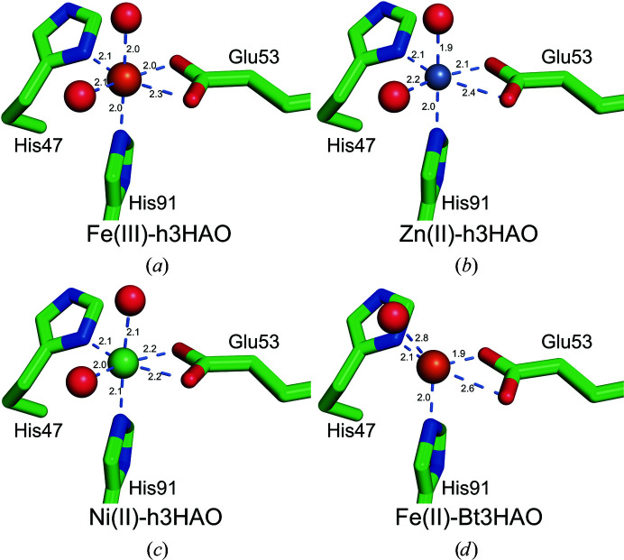
Figure 6.
Comparison of the metal–ligand distances for all of the available mammalian 3HAO crystal structures: Fe(III)-h3HAO (this work; PDB entry 5tk5), Zn(II)-h3HAO (this work; PDB entry 5tkq), Ni(II)-h3HAO (PDB entry 2qnk) and bovine 3HAO [Fe(III)-Bt3HAO; Đilović et al., 2009 ▸]. Fe atoms are shown as orange spheres, the Ni atom is shown as a green sphere and the Zn atom is shown as a gray sphere. Metal–ligand interactions are indicated by light blue dashed lines.

