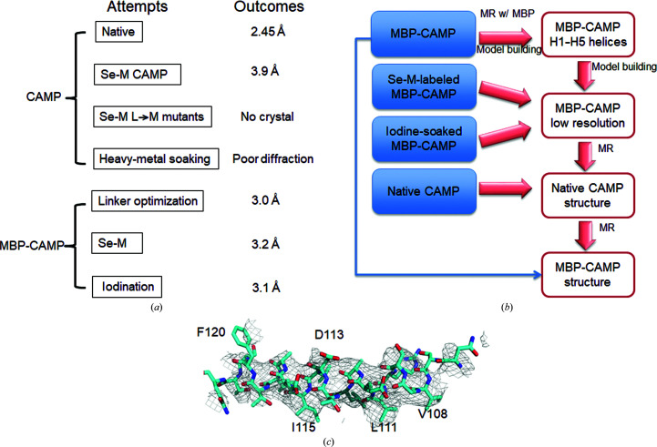
Figure 2.
A flowchart for the structural determination of CAMP factor. (a) Data sets collected to solve the CAMP structure. Seven types of protein crystals were tested for X-ray diffraction and data collection. (b) A flowchart describing CAMP structure determination. Native MBP-CAMP fusion crystals diffracted to 3.0 Å resolution and the structure was solved by molecular replacement using MBP as a search model. The heavy atoms introduced by Se-M labeling and iodine soaking also provided guidance for manual model building and side-chain assignment of the CAMP region. (c) Partial difference electron-density map from MBP-CAMP data at 3.0 Å resolution after molecular replacement with MBP. The F o − F c map of the H5 region is contoured at 1σ. The map is superimposed with the final model of H5 (cyan).

