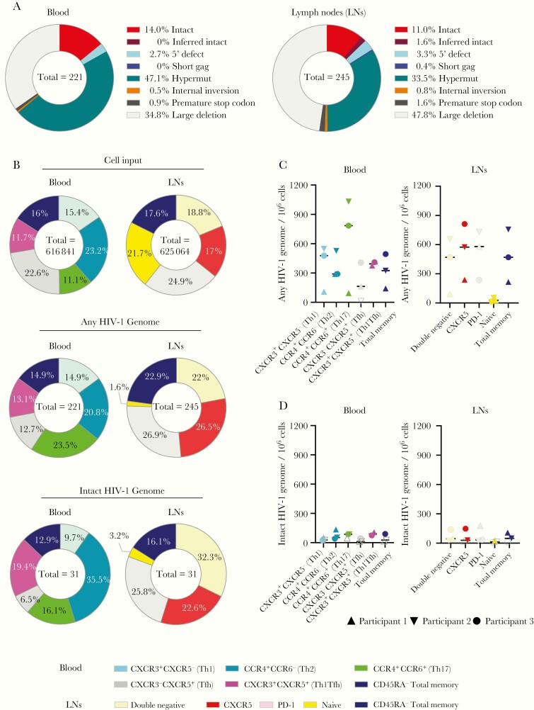
Figure 1.
Frequency of intact and defective human immunodeficiency virus (HIV) 1 sequences in CD4 T-cell subsets from peripheral blood and lymph node samples from antiretroviral-treated persons with HIV-1. A, Pie charts reflecting the relative proportion of intact and defective HIV-1 sequences isolated from lymph node and blood of the 3 study participants with HIV-1. B, Pie charts indicating the relative contribution of phenotypically distinct CD4 T-cell populations to the total number of cells analyzed (upper panels) and to the total number of any or intact HIV-1 sequences (lower panels) in blood and tissues. C, D, Frequencies of any HIV-1 sequences (C) or intact HIV-1 sequences (D) derived from indicated subsets of CD4 T cells from blood or lymph node compartments. Open symbols represent data below the limit of detection, calculated by assuming the presence of 0.2 viral copies in the number of cells in which no target was identified, and bars represent median values. Abbreviations: PD-1, programmed cell death 1; Tfh, T follicular helper; Th1, Th2, and Th17, T-helper 1, 2, and 17.

