Figure 1.

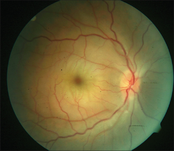
Fundus picture of the right eye showing the cherry red appearance of the fovea and opaque edematous macula. Arteries are significantly narrowed, and there is some disc swelling

Fundus picture of the right eye showing the cherry red appearance of the fovea and opaque edematous macula. Arteries are significantly narrowed, and there is some disc swelling