
Fig. 2. Sirt7−/− females contain fewer gametes.
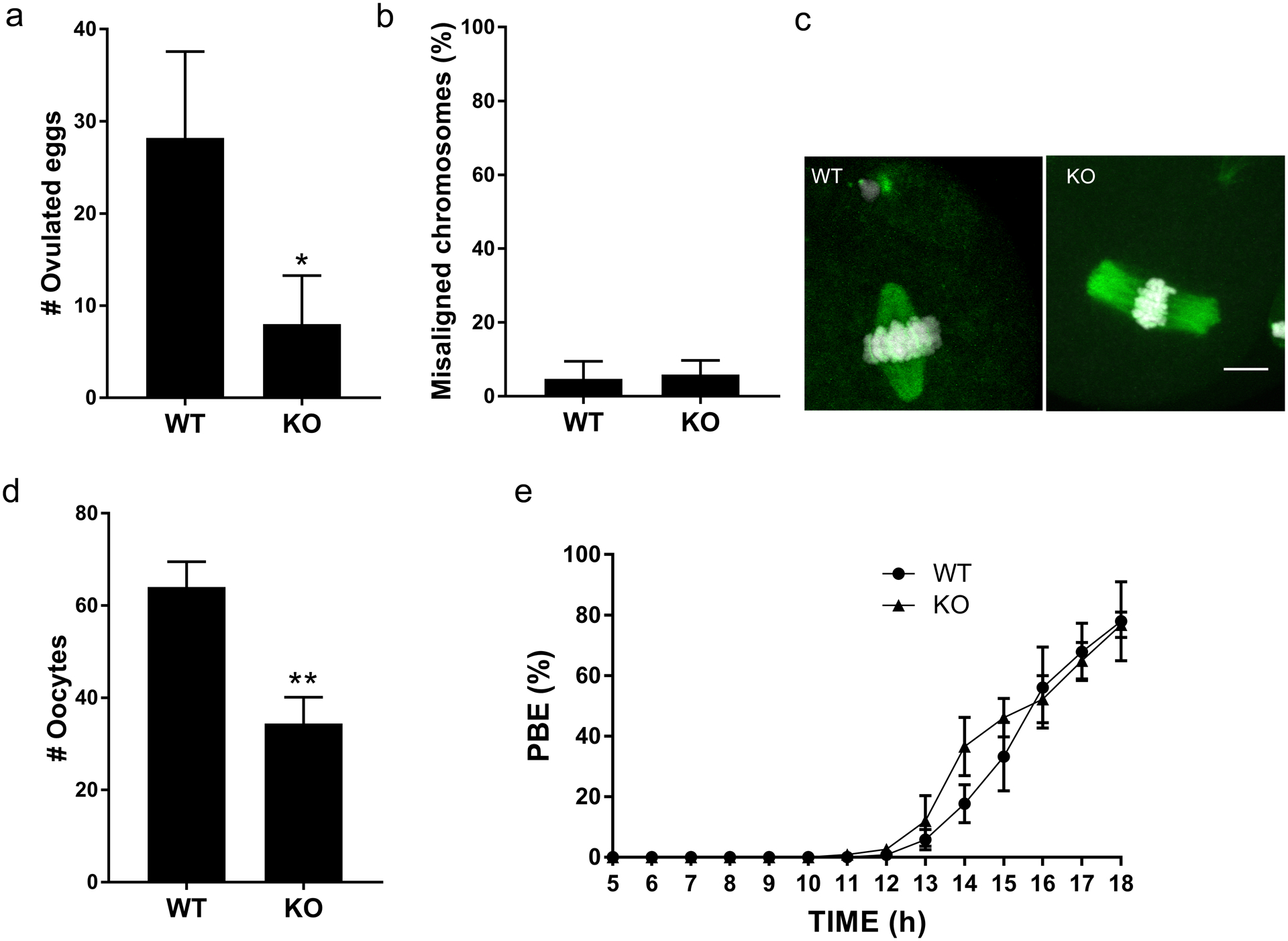
(a) Number of ovulated eggs isolated after hormone stimulation from wild-type (WT) and Sirt7−/− (KO) females. Graph represents the mean ±SEM (n=5 mice /genotype). (b) Percent of misaligned chromosomes from ovulated eggs. Data represents the mean ±SEM (n=3 animals/per genotype; ~40 eggs were evaluated). (c) Representative images of WT and KO eggs stained to detect DNA (DAPI, white) and the spindle (α-tubulin, green). (d-e) Prophase I-arrested oocytes from WT and KO animals were collected (d) and in vitro matured on a live-cell imager for 18h and the time of polar body extrusion (PBE) (e) was measured. Graph represents mean ±SEM (n=3 animals/genotype). * P< 0.05; ** P < 0.01; scale bar, 10μm